What Does Your Prostate Size Actually Tell Your Doctor?
Most patients pay close attention to their PSA level. They remember the number, track whether it rises or falls, and compare it to previous results.
What many patients don’t realize is that their physician may also be paying close attention to another number: the size of the prostate itself.
Prostate size rarely receives the same attention as PSA, MRI findings, or biopsy results. Yet it can influence how doctors interpret screening tests, evaluate symptoms, plan biopsies, and even consider treatment options.
In other words, prostate size is not simply an anatomical detail. It is a piece of clinical information that helps place many other findings into context.
1. Prostate Volume Is More Than a Measurement
When physicians talk about prostate size, they are usually referring to prostate volume. Volume describes the three-dimensional size of the gland rather than just its length or width.
This matters because the prostate is not a fixed-size organ. Some men have relatively small prostates throughout their lives. Others develop significantly larger glands as they age. Two patients may have identical PSA levels, identical symptoms, and identical ages, yet have very different prostate volumes.
That difference can influence how physicians interpret nearly every other piece of information they collect.
One of the most common reasons for prostate enlargement is a condition called Benign Prostatic Hyperplasia (BPH). BPH is not cancer. It refers to a non-cancerous increase in the number of prostate cells that occurs in many men as they age.
As the prostate enlarges, it may contribute to symptoms such as:
- Frequent urination
- Weak urinary stream
- Difficulty emptying the bladder
- Increased nighttime urination
However, BPH also creates another challenge. A larger prostate contains more prostate tissue and more prostate tissue often produces more PSA. This is one reason physicians rarely evaluate PSA without considering prostate size at the same time.
Small differences in volume measurements can influence clinical calculations and decision-making, so how the volume is determined matters.
Historically, physicians estimated prostate size using physical examination or ultrasound. These methods can be helpful, but they are not always precise. MRI provides a more detailed view of the gland, allowing radiologists to measure its length, width, and height and calculate an overall volume.
It's worth understanding that this calculation is still an estimate. It uses the ellipsoid formula — length × width × height × 0.52 (an approximation of π/6) — which assumes the prostate is a smooth, symmetrical, egg-like shape. Real prostates aren't perfectly shaped that way, so the formula often undercounts the true volume.
AI can take this a step further. Instead of relying only on three measurements, some AI systems can trace the gland’s outline on individual MRI slices and combine those outlines into a three-dimensional, segmentation-based volume estimate. This approach follows the prostate’s actual shape more closely than the ellipsoid formula, which can be especially helpful when the gland is irregularly shaped.
Like any imaging-based measurement, this is still an estimate and can depend on image quality, software performance, and how clearly the prostate boundaries can be identified. But compared with a simple length × width × height calculation, segmentation can provide a more detailed view of prostate volume.
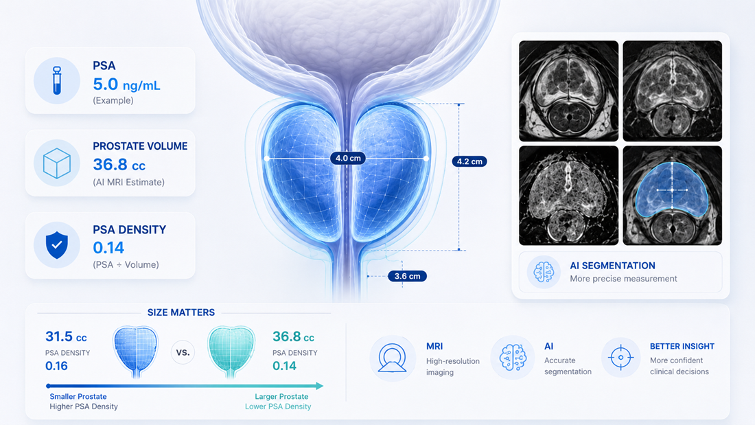
For example, a prostate measuring 4.0 × 3.6 × 4.2 cm gives an ellipsoid estimate of:
4.0 × 3.6 × 4.2 × 0.52 ≈ 31.5 cc
Measuring the same gland slice by slice, AI finds the true volume is closer to 36.8 cc — about 17% larger. That difference may seem minor, but it can change other calculations physicians rely on, as the next section shows.
A PSA level never exists in isolation. Physicians often ask, "Is this PSA appropriate for a prostate of this size?" That question leads to a measure called PSA density, which compares the PSA level to the volume of the prostate. The same PSA number can mean something quite different in a small prostate than in a large one — the number hasn't changed, but the context has.
This is exactly why an accurate volume matters. Returning to the prostate from the previous section, imagine this patient has a PSA of 5.0 ng/mL:
Using the ellipsoid estimate of 31.5 cc, the PSA density is 5.0 ÷ 31.5 ≈ 0.16 -- above the commonly used threshold of about 0.15, which would typically raise concern.
Using the AI's more accurate volume of 36.8 cc, the PSA density is 5.0 ÷ 36.8 ≈ 0.14 -- below that threshold, and more reassuring.
Same patient, same PSA, but a more accurate volume may shift the result from concerning to reassuring. That kind of difference can influence whether a patient is advised to proceed to a biopsy or to continue monitoring, which is why precise volume assessment has become an increasingly valuable part of modern prostate MRI evaluation.
Once a biopsy is recommended, prostate size matters again though this time in how the procedure is carried out.
A larger gland contains more tissue, and as the prostate grows, it can become more challenging to sample every region thoroughly. The same number of biopsy needles covers a smaller fraction of a large gland than a small one, so physicians weigh anatomy, lesion location, and gland size when planning how a biopsy should be performed.
This is where MRI is especially valuable, because it helps identify:
- Suspicious lesions
- Lesion location
- The relationship between lesions and surrounding anatomy
- Overall gland volume
Modern prostate biopsies increasingly use this information to target specific areas rather than sampling the gland blindly. AI can support this step by highlighting suspected regions of concern and marking their location on the MRI with color overlays, giving the radiologist and treating physician a clearer map of where to focus. This helps the physicians planning the procedure to see the most relevant areas as clearly as possible.
The goal is not simply to perform a biopsy. It's to gather the most meaningful information possible from the procedure.
Prostate volume is not just another number in an MRI report. It influences how physicians interpret PSA levels, calculate PSA density, evaluate risk, and plan biopsies. Because so many important decisions can depend on these measurements, accuracy matters.
This is one reason some patients seek a second review of their prostate MRI.
A second review may provide additional insight into findings that can influence clinical decision-making, including prostate volume, lesion characteristics, lesion location, and other imaging features that may affect how the scan is interpreted.
In recent years, artificial intelligence (AI) has become another tool that can assist in this process. AI systems can analyze large amounts of imaging data, perform quantitative measurements, and identify patterns that may not be immediately obvious during a routine review.
Services such as DeepView Imaging provide advanced AI analysis of a prostate MRI, giving patients and their care team an additional perspective on the information already contained within a prostate MRI.
The goal is not to replace the radiologist or treating physician. The goal is to help patients and physicians make important decisions with the most complete information possible.
Disclaimer: This article is educational, isn't medical advice, and decisions should be made with your treating physician.