Can a Prostate MRI Miss Cancer?

"Can an MRI miss prostate cancer?" This is one of the most unsettling questions you may quietly ask after receiving MRI results, particularly if your PSA levels remain elevated or your doctor continues recommending monitoring after a “normal” MRI.
Most people assume a normal prostate MRI completely rules out cancer. In reality, prostate MRI is one of the most advanced tools available for evaluating prostate cancer risk, but it still has limitations that many patients are never fully told about.
A prostate MRI does not simply answer: “Is cancer there or not?”
Instead, it helps doctors estimate how suspicious a finding may be, how aggressive it could appear, and whether further testing is necessary.
Understanding how MRI actually works, what it is good at detecting, and where uncertainty can still exist can help you feel more informed and less overwhelmed when making decisions about biopsy, monitoring, or next steps.
What a Prostate MRI Is Actually Looking For
One of the biggest misconceptions about prostate MRI is that it works like a photograph where cancer either clearly appears or does not.
In reality, radiologists are analyzing patterns inside the prostate that may suggest abnormal tissue behavior. A prostate MRI evaluates how tissue looks, how dense it appears, how water molecules move through it, how blood flows through certain areas, and whether suspicious tissue behaves differently than normal prostate tissue across multiple imaging sequences.
This is the value of a prostate MRI. Doctors are not relying on a single image. They are combining several different types of imaging information together to estimate how concerning a finding may be.
Some cancers create very obvious changes and stand out clearly. Others do not. More aggressive tumors are often easier to detect because they disrupt surrounding tissue in more visible ways. Smaller or slower-growing cancers may create much subtler changes that are harder to distinguish from normal tissue.
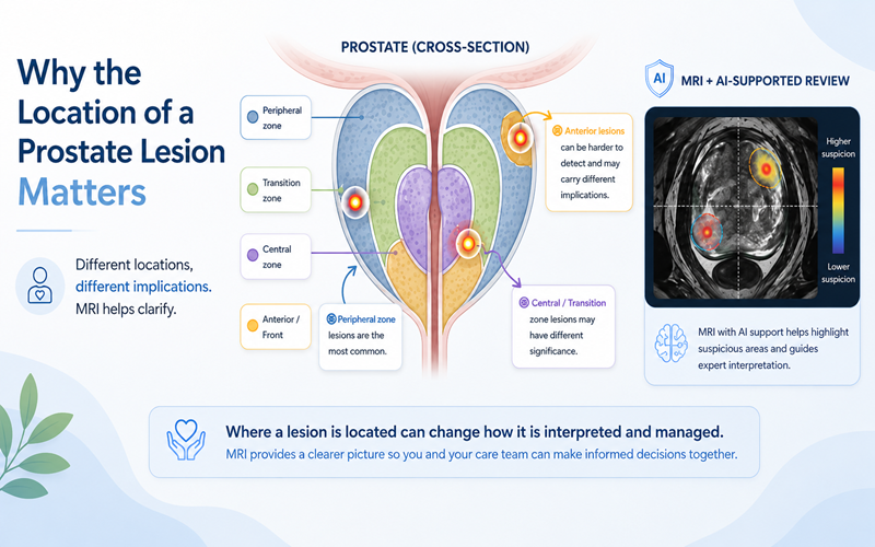
Location also matters. Certain parts of the prostate are more difficult to evaluate than others, and some abnormalities can blend into surrounding tissue in ways that make interpretation challenging.
This is one reason prostate MRI is so valuable, but also why it cannot provide absolute certainty in every case.
If your MRI report says phrases like “low suspicion” or “no clinically significant cancer identified,” you may naturally feel relieved. But these phrases are also commonly misunderstood.
In prostate imaging, the term “clinically significant” has a very specific meaning. It usually refers to cancers that are more likely to grow, spread, or eventually require treatment.
So when an MRI report says no clinically significant cancer was identified, it often means the scan did not show strong evidence of a more aggressive or clearly visible cancer.
That does not necessarily mean no cancer exists at all.
In fact, one of the major goals of prostate MRI is to help distinguish between cancers that may require treatment and slower-growing cancers that may never become dangerous during your lifetime. This is important because many prostate cancers grow very slowly, and overtreatment has become a major concern in prostate cancer care.
That is one reason your doctor may still recommend monitoring even when there is still some uncertainty. The goal is not simply to find every possible abnormal cell. The goal is to identify cancers that are truly likely to affect long-term health while avoiding unnecessary procedures and side effects whenever safely possible.
For patients, understanding this distinction can make MRI results feel less confusing and more logical.
If you want a deeper explanation of MRI terminology, lesions, and PI-RADS scoring, you can also read our guide:
“How to Read Your Prostate MRI Results”
One of the most frustrating situations is when your PSA levels remain elevated even though the MRI appears reassuring.
Naturally, you may begin wondering which result you are supposed to trust.
The important thing to understand is that PSA and MRI measure very different things. PSA is a blood marker that reflects activity happening somewhere in the prostate, but it does not specifically identify where that activity is coming from. PSA can rise because of cancer, but it can also increase due to inflammation, infection, enlargement of the prostate, recent procedures, or even normal variation over time.
MRI works differently. Instead of measuring prostate activity broadly, it looks for visible structural or functional abnormalities inside the gland itself.
Because these tests measure different things, they do not always perfectly align. You can have an elevated PSA with a reassuring MRI, or a concerning MRI finding with only mildly elevated PSA levels.
This is why experienced doctors rarely rely on a single test in isolation. Instead, they look for patterns over time and combine multiple pieces of information together before recommending biopsy, monitoring, or treatment.
Understanding this can help you realize that differing results do not automatically mean something was missed or that the testing failed.
You may assume prostate MRI interpretation is completely objective, as if every radiologist reviewing the same scan would automatically reach the same conclusion.
In reality, prostate MRI interpretation is highly specialized.
Some findings are obvious. Others fall into gray areas where experience matters significantly. Small lesions, borderline abnormalities, or PI-RADS 3 findings can sometimes be interpreted differently depending on the radiologist’s training, how often they read prostate MRIs, and their level of specialization in prostate imaging.
This does not mean MRI is unreliable. It means that medicine often involves interpretation and judgment, especially in borderline cases.
This is also one reason second opinions have become increasingly common in prostate imaging. In some situations, another specialist may interpret a subtle finding differently or provide additional confidence about the original assessment.
Newer technologies such as AI-supported analysis are also being explored as ways to improve consistency and help identify subtle imaging patterns that may be difficult to detect visually alone.
If your doctor recommends continued monitoring instead of rushing directly to biopsy, it can sometimes feel unsettling. You may even wonder if “waiting” means something is being missed.
But in many situations, monitoring is actually a very intentional medical strategy.
Doctors are constantly balancing two important goals at the same time: avoiding missed aggressive cancer while also avoiding unnecessary biopsies and overtreatment.
Because prostate biopsies carry risks and because many prostate cancers grow very slowly, doctors often look carefully at your overall risk picture before recommending invasive procedures.
This is where MRI has become especially valuable. A reassuring MRI may help support a decision to monitor PSA trends, repeat imaging later, or continue surveillance rather than immediately moving to biopsy.
For many men, this can be one of the hardest emotional parts of the process. Monitoring can feel uncertain because there is no immediate “solution” or definitive answer. But in many cases, careful monitoring is not passive. It is a medically informed approach designed to avoid unnecessary treatment while still watching closely for meaningful changes over time.
One of the hardest parts of prostate cancer evaluation is realizing that uncertainty can still exist even after advanced testing.
You may have gone into the MRI hoping for a definitive answer. When the results still leave room for interpretation, it can create anxiety, frustration, and decision fatigue.
But uncertainty does not automatically mean danger.
In reality, many prostate cancer decisions are not about achieving perfect certainty. They are about gathering enough reliable information to make the safest and most informed next step possible.
A prostate MRI remains one of the most important advances in prostate cancer detection and risk assessment. It has helped many men avoid unnecessary biopsies while improving the ability to identify more concerning cancers earlier.
If your MRI results still feel unclear, you are not alone. Many patients seek additional clarity before making decisions about biopsy, monitoring, or treatment.
In some situations, that may involve another specialist reviewing the scan or newer approaches such as AI-supported analysis. Some patients choose services like DeepView Imaging as part of that process.
The goal is not to create more fear. The goal is to help you feel more informed, more confident, and better supported as you move forward.
Frequently Asked Questions About Prostate MRI
Can a prostate MRI miss cancer?
Yes. While prostate MRI is one of the best tools available for evaluating prostate cancer risk, some cancers can still be difficult to detect, especially if they are very small, low-grade, or located in areas that are harder to evaluate clearly on imaging.
Can PSA still be high if the MRI looks normal?
Yes. PSA and MRI measure different things. PSA reflects activity happening somewhere in the prostate, while MRI looks for visible abnormalities inside the gland itself. PSA can also rise because of inflammation, infection, or benign enlargement of the prostate.
Does a “normal” MRI mean I definitely do not have cancer?
Not necessarily. A reassuring MRI means no clearly visible signs of clinically significant cancer were identified, but smaller or lower-grade cancers may not always appear clearly on imaging.
What does “clinically significant cancer” mean?
Clinically significant cancer usually refers to cancers that are more likely to grow, spread, or require treatment. Many slower-growing prostate cancers may never become dangerous during a patient’s lifetime.
Why might two doctors interpret the same MRI differently?
Prostate MRI interpretation is highly specialized. Small lesions, PI-RADS 3 findings, and subtle abnormalities can sometimes be interpreted differently depending on a radiologist’s experience and specialization in prostate imaging.
Why would my doctor recommend monitoring instead of immediate biopsy?
In some situations, monitoring is the safest and most appropriate approach. Doctors try to balance detecting aggressive cancer early while also avoiding unnecessary biopsies and overtreatment for slower-growing disease.