
Can AI Help Read Your Prostate MRI? Why Training Data Matters
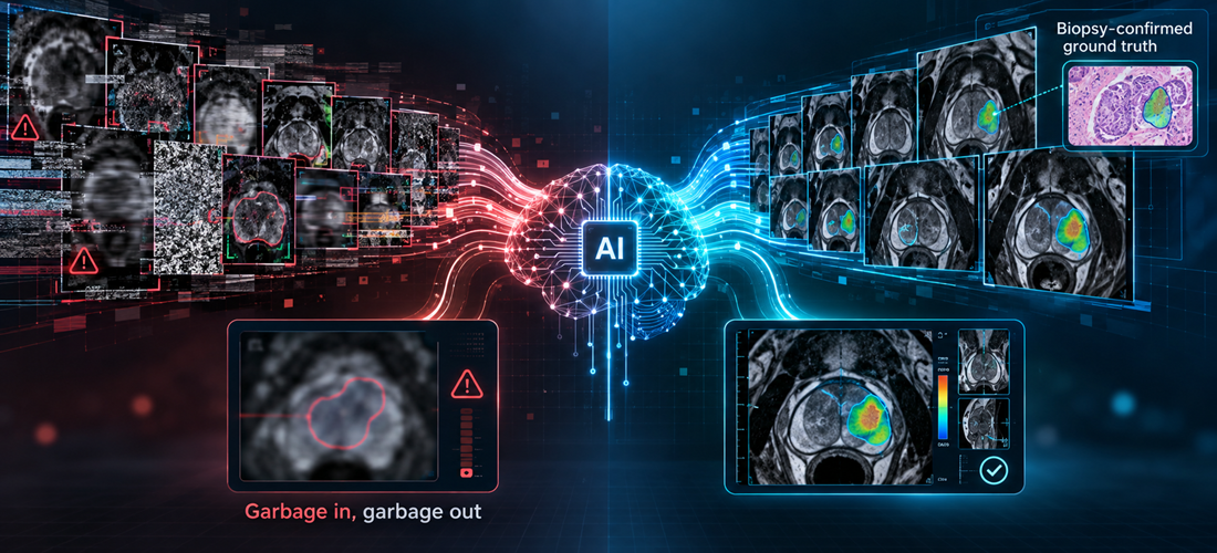
If you've had a prostate MRI, you may have heard that artificial intelligence (AI) can help review it. That raises two fair questions. Does AI actually add anything? And how can you tell whether a particular AI tool is reliable? The second answer depends largely on what the AI learned from and how it was tested.
Why use AI to help read a prostate MRI?
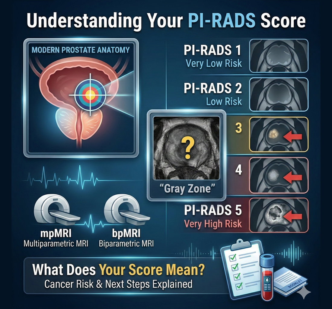
A radiologist reviews several sets of MRI images and scores any suspicious area, usually with the Prostate Imaging Reporting and Data System (PI-RADS), a scale from 1 to 5. In the UK, NICE guidance recommends a similar five-point Likert scale. A PI-RADS score describes how suspicious an area appears on MRI; it is not the same as a diagnosis.
Reading these scans takes skill, and radiologists do not always agree. A 2022 review found substantial agreement overall, with more disagreement around borderline findings such as PI-RADS 3. MRI interpretation can vary because scan quality, lesion location, reader experience, and clinical context all matter.
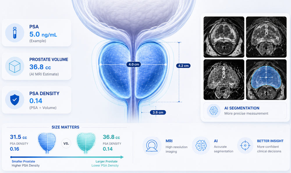
AI may help in several ways. It applies the same approach to every scan and does not get tired. It can outline the prostate and any suspicious areas, estimate prostate volume, and point to regions that deserve a closer look.
The research is encouraging, and well-designed studies rely on exactly the kind of data this article describes. In the international PI-CAI study, published in 2024, an AI system learned from more than 9,000 prostate MRI exams, with cancer-positive cases confirmed by biopsy. It was then tested on 1,000 scans it had never seen, where significant cancers were confirmed by biopsy or surgery and cases without significant cancer were checked with at least three years of follow-up.
Compared with 62 radiologists reading the same 400 scans, the AI detected the same share of significant cancers while raising about half as many false alarms. Its performance was also very close to the original clinical reports, which drew on each patient’s history and team discussion, although it fell short of the study’s pre-set statistical test for showing it performed at least as well. The authors called for further testing in everyday care.
Results like these apply to the system that was tested, which is why every AI tool needs its own evidence. ProstatID’s™ testing is described later in this article. AI can still miss cancer or flag harmless tissue, so its best-supported role is as a second set of eyes alongside a physician.
AI learns by studying examples. A prostate MRI training set may include:
- MRI images. T2-weighted images show anatomy. Diffusion-weighted images (DWI) and apparent diffusion coefficient (ADC) maps show how freely water moves through tissue, which often changes where cancer is present.
- Expert markings. Specialists outline the prostate and any suspicious regions.
- Biopsy findings. Tissue samples examined by a pathologist show whether a sampled area was benign or cancerous.
From these examples, the system learns patterns linked to clinically significant prostate cancer, meaning cancer more likely to grow and spread (usually Grade Group 2 or higher). Lower-grade cancers are often monitored through active surveillance rather than treated right away.
Some scans also include images taken after a contrast injection, called dynamic contrast enhancement (DCE). Different AI tools use different image types.
Biopsy links are what make training data especially valuable. An AI trained mainly on what radiologists thought looked suspicious learns to copy their impressions, uncertainty included. One trained on scans linked to biopsy results learns from what was confirmed in tissue, especially when each marked spot is tied to a specific biopsy sample. Biopsies can miss cancer too, so even carefully verified data are imperfect, and diagnosis still depends on pathology.
Before training, specialists check scan quality, confirm pathology results, and mark the prostate and any lesions. Inconsistent labels can teach a system misleading patterns.
As an illustration, using hypothetical numbers: 2,000 scans with reliable biopsy-linked findings and careful expert markings might teach a system more than 10,000 scans with inconsistent labels and uncertain outcomes. Size helps, but more data is not automatically better data.
Variety matters too. Prostate MRIs are performed on scanners with different magnet strengths, mainly 1.5 tesla (1.5T) and 3 tesla (3T), from different manufacturers. Both can produce useful prostate images, yet the pictures look subtly different. An AI trained and tested on older and newer equipment is better prepared for the range of scans patients actually receive, although variety alone does not guarantee accuracy.
A system should be tested on cases that were set aside before training and never used to build or adjust it. Comparing the AI's results on these held-out cases with biopsy or follow-up findings shows whether it learned patterns that carry over to new patients or simply memorized its examples.
Reader studies, in which physicians interpret the same scans with and without AI, show whether it helps the people using it. Most of these studies look back at past scans, so they cannot yet show whether AI improves long-term patient outcomes.
In the United States, the Food and Drug Administration (FDA) reviews this kind of evidence before clearing medical AI software for a defined use. The EU and UK use separate pathways, including CE and UKCA marking. Clearance shows a tool met regulatory standards for its intended use, though no tool is right every time.
General-purpose chatbots such as ChatGPT and Claude are large language models (LLMs), trained mostly on huge amounts of text. They can help explain terms in your MRI report and prepare questions for an appointment. Some can look at images, yet they were not trained on biopsy-linked prostate MRI, have not been tested on held-out prostate cases, and are not cleared to interpret prostate MRI or diagnose cancer.
A prostate MRI also contains many images across several sequences, so a few screenshots leave out most of the information. And chatbots can confidently describe abnormalities that are not there. A 2024 study of an earlier image-capable version of GPT-4 on emergency CT, X-ray, and ultrasound images found it identified the correct finding only about a third of the time and described problems that were not present in nearly half of cases. It did not test prostate MRI or today's newer models, but it shows why general image-reading ability differs from validated prostate MRI analysis.
If you use a chatbot to understand a written report, check its privacy terms first.
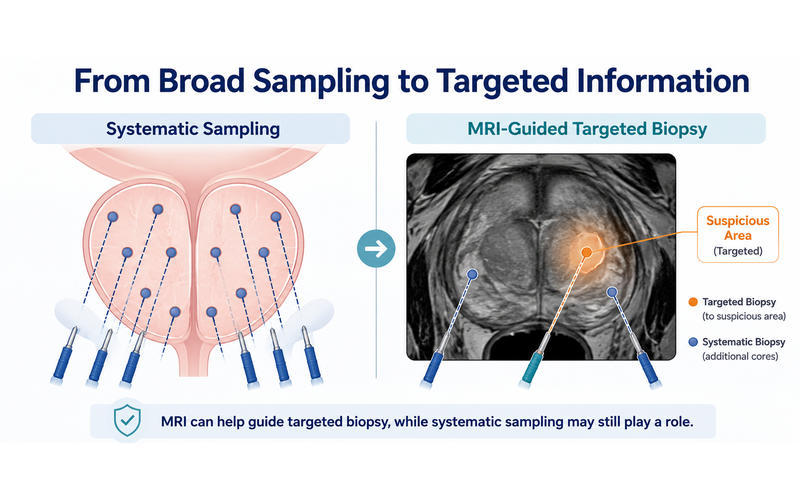
Prostate MRI helps identify suspicious areas, guide decisions about targeted biopsy, and support monitoring. A negative MRI lowers risk, but it does not reduce the risk to zero. An AI-supported review may add lesion outlines, measurements, and risk scores, yet it cannot decide on its own whether you have cancer.
Biopsy decisions often depend on the full clinical picture: your prostate-specific antigen (PSA) level and trend, PSA density (PSA relative to prostate volume), MRI score and lesion location, age, family history, ancestry where relevant, prior biopsies, symptoms, other health conditions, and personal preferences. Similar numbers can mean different things for different people.
If an AI-highlighted area does not appear in your original report, that does not automatically mean cancer was missed. Any difference is worth discussing with your physician in context.
Questions worth asking about any AI tool:
- Was it trained on biopsy-confirmed cases marked by experts?
- Was it trained and tested on different types of MRI scanners?
- Has it been tested on patients outside its training data?
- Is it cleared or certified for its stated use where I live?
- How should its findings be read alongside my MRI report and other results?
Asking the same questions of ProstatID
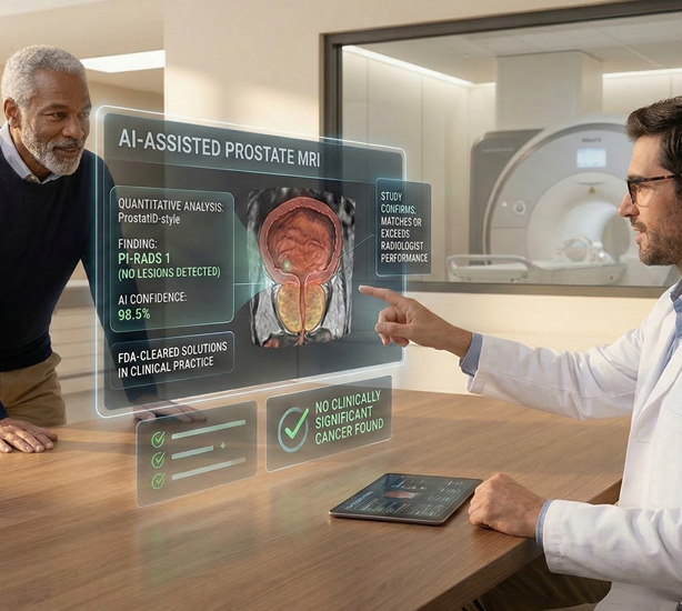
These questions apply to any AI tool, including the one DeepView Imaging uses. DeepView offers an AI-supported second analysis of an existing prostate MRI using ProstatID, software from Bot Image. Here is how it answers them:
- Biopsy-confirmed, expert-marked data? According to Bot Image, ProstatID learned from more than 6,000 biopsy-confirmed data points across over 1,000 cases. Each data point links a spot on the MRI to a biopsy result, and all were curated and marked by prostate imaging experts.
- Different scanners? Its training included both 1.5T and 3T systems, and it is cleared for use with scanners from several major manufacturers.
- Tested on patients it had not seen? For its FDA review, itwas tested on cases set aside before training. It was also evaluated in a study of nine physicians who read the same scans with and without it. Like most AI studies, this testing looked back at past scans.
- Cleared where you live? ProstatID is FDA-cleared in the U.S., CE-Marked in the EU, and UKCA-Certified in the UK as an aid to physicians reading prostate MRI. It analyzes T2-weighted, DWI, and ADC images, so scans with or without contrast can be used.
- How should the results be read? Alongside your original report, with your physician. The DeepView report shows color overlays and risk scores to discuss at your next appointment. It does not diagnose cancer or replace your radiologist, urologist, or treating physician.
AI can add a consistent second look at a prostate MRI, and research suggests it may help. How much you can rely on a particular tool depends on what it learned from and how it was tested: biopsy-confirmed examples, expert annotation, varied scanners, and independent validation. No AI result confirms or rules out cancer on its own, but understanding how a tool was built can help you have a more informed conversation with your physician.
This article is for educational purposes only and is not medical advice. Decisions about screening, biopsy, diagnosis, monitoring, or treatment should be made with your treating physician.
Sources
- Saha A, Bosma JS, Twilt JJ, et al. Artificial intelligence and radiologists in prostate cancer detection on MRI (PI-CAI): an international, paired, non-inferiority, confirmatory study. The Lancet Oncology. 2024;25(7):879–887.
- Wen J, Ji Y, Han J, Shen X, Qiu Y. Inter-reader agreement of PI-RADS v2.1: A systematic review and meta-analysis. Frontiers in Oncology. 2022;12:1013941.
- Turkbey B, Rosenkrantz AB, Haider MA, et al. PI-RADS Version 2.1: 2019 Update. European Urology. 2019;76(3):340–351.
- Brin D, Sorin V, Barash Y, et al. Assessing GPT-4 multimodal performance in radiological image analysis. European Radiology. 2025;35(4):1959–1965 (online 2024).
- American Urological Association / Society of Urologic Oncology. Early Detection of Prostate Cancer: Guideline Part II, Considerations for a Prostate Biopsy. The Journal of Urology. 2023.
- National Institute for Health and Care Excellence (NICE). NG131: Prostate cancer, diagnosis and management. Published 2019; updated 2021.
- U.S. Food and Drug Administration. ProstatID 510(k) clearance, K212783. July 8, 2022.
- Bot Image, Inc. ProstatID manufacturer information. Used only for product description, including training data figures; manufacturer statements are not independent clinical evidence.
- Bot Image, Inc. ProstatID CE marking announcement. October 27, 2025; UKCA certification announcement. November 18, 2025. Manufacturer announcements.
More Articles

.svg)

