September is Prostate Cancer Awareness Month.
Learn the facts and understand your options.
Learn the facts and understand your options.

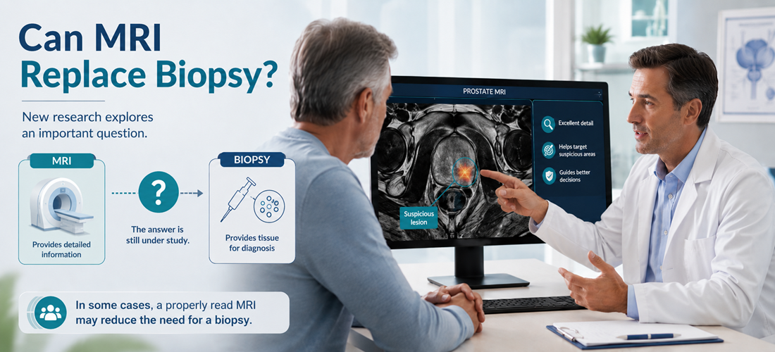
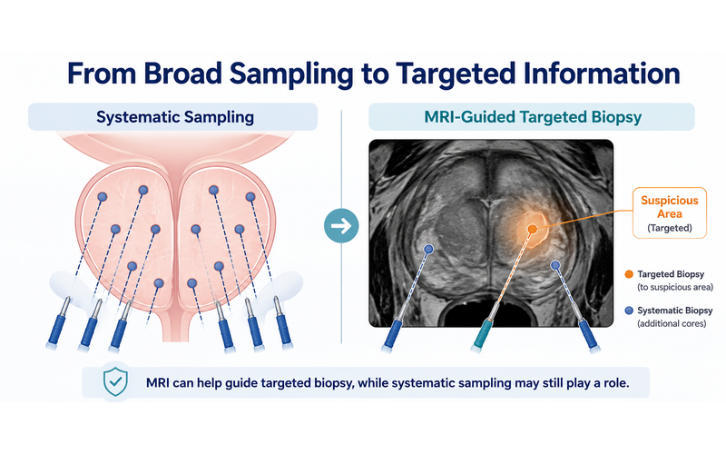
Waiting for a prostate biopsy is one of the harder stretches of the diagnostic pathway. The MRI is done, something suspicious has been seen, and the answer that matters most still sits weeks away behind another procedure. A study published in the European Journal of Radiology by a team at University College London (UCL) and University College London Hospitals (UCLH) asks a question many men in that position have wondered about: how much can the scan itself already tell us?
For some cancers, imaging alone is enough to begin treatment. Kidney and testicular tumors are often treated without a biopsy first. Prostate cancer has not worked that way, because MRI has been understood as a tool for finding suspicious areas rather than confirming what they are. This research explores whether that line might sit slightly differently for a small group of men.
The short answer, for today, is that biopsy remains the standard. The more interesting answer is that for a minority of men, the scan may already be carrying more certainty than we have been giving it credit for.
The team looked at 880 consecutive men who attended a one-stop prostate clinic at UCLH over one year and had a prostate MRI. Of those, 251 had scans read at the time as moderately or highly suspicious for cancer.
Four experienced radiologists re-read all 251 scans independently, at least three years later, blinded to the biopsy results. They knew each man's age and PSA (prostate-specific antigen, a blood protein that can rise with cancer and with benign conditions), and nothing else. For each scan they recorded one of three levels of confidence that clinically significant cancer was present: "99% sure," "90% sure," or "less than 90% sure." Where they disagreed, they discussed the case and reached a consensus.
Clinically significant cancer here means Gleason Grade Group 2 or higher, the type more likely to grow and spread, as opposed to low-grade disease that is often monitored rather than treated.
In the consensus read, 79 men fell into the "99% sure" group. Sixty-nine went on to targeted biopsy, and all 69 had clinically significant cancer. Nearly three-quarters had Grade Group 3 or higher. A further 45 men were rated "90% sure"; of the 41 biopsied, 40 had significant cancer, and the one man who did not had low-grade Grade Group 1 disease.
In the "less than 90% sure" group, 42% of those biopsied had significant cancer, and only 7% had Grade Group 3 or higher. The readers agreed with one another at a level the authors describe as substantial.
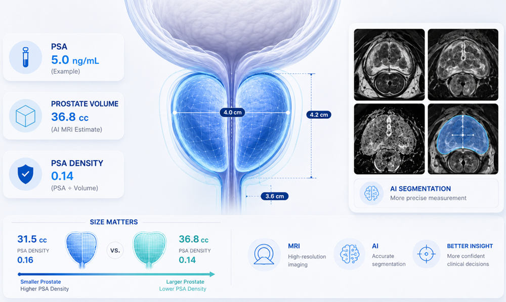
One detail stands out. Confidence did not depend on the tumor being large or the PSA being alarming. In the "99% sure" group, about a third of tumors were under 1 cc and about a quarter of the men had a PSA density below 0.15 ng/ml/ml, a figure often read as reassuring. One case in the paper involved a 65-year-old man with a PSA in the normal range for his age whose small lesion was called with high confidence by all four readers and proved to be Grade Group 3 cancer at biopsy.
The authors are careful about the limits of the work, and those limits matter.
This was a single specialist referral center, and all four readers were highly experienced, with 5 to 23 years of prostate MRI reporting behind them. The paper says plainly that the findings may not carry over to settings with lower scan volumes or less specialised readers, and describes itself as hypothesis-generating, with multi-center validation required before anything changes in practice.
Not every man in the study was biopsied, so the histology picture is incomplete, and the scans used contrast, which is not universal practice. The certainty being described also runs in one direction only. A confident MRI read may point strongly toward cancer, while a reassuring MRI still does not rule cancer out; a negative or low-suspicion MRI lowers risk without reducing it to zero.
Nothing here suggests a man should skip a recommended biopsy. Diagnosis still rests on tissue, and the pathology report carries grading information that guides treatment planning.
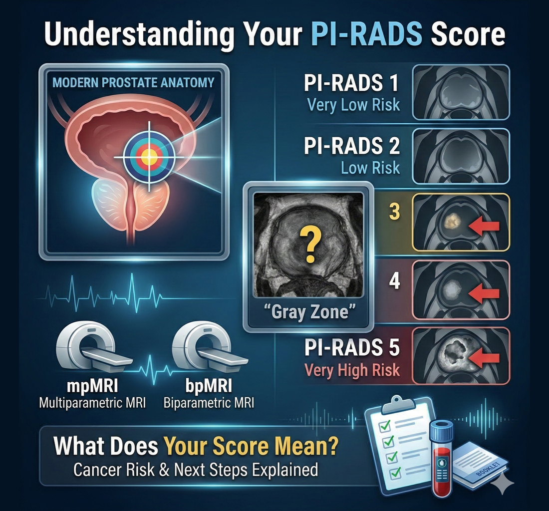
The researchers deliberately stepped outside PI-RADS, the standard five-point system used to describe how suspicious an area looks on prostate MRI. In the UK, NICE guidance also permits a five-point Likert scale, which is what UCLH uses. Their reasoning was that PI-RADS was designed to decide who needs a biopsy, and it does that well. It was not built to single out the scans where cancer is close to certain. Published false-positive rates for a PI-RADS 5 score sit around 17%, too high to act on without confirmation.
For patients, the practical point is this. A PI-RADS or Likert score describes how suspicious an area appears; it is not the same as a diagnosis, and two radiologists looking at the same images can reach different scores. Scan quality, lesion location, reader experience and clinical context all feed into the result.
For more information see: Understanding Your PI-RADS Score
The UCL findings rest on something specific: four experienced readers looking carefully at the same images, then talking through their disagreements. The depth and repetition of the review mattered as much as the scan itself. Most men do not get four readers and sometimes the one they do get has less experience reading prostate MR ... another reason is why a second-opinion is important.
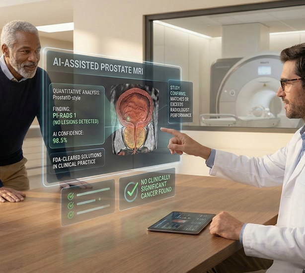
DeepView Imaging provides an AI-supported second analysis of a prostate MRI a man has already had, using ProstatID™, AI software from Bot Image that is FDA-cleared, CE-Marked and UKCA-Certified. Its FDA clearance covers use as both a detection (CADe) and diagnostic (CADx) aid, in patients undergoing high-risk screening as well as diagnostic prostate MRI. The software reads the T2-weighted, diffusion-weighted and ADC sequences, so it works with biparametric and multiparametric scans alike, and returns lesion segmentation and a risk score shown as a colour overlay. The goal is to give patients and their care team another perspective on information already contained within the MRI, which can be shared with a physician when discussing next steps. It does not replace a radiologist, urologist or treating physician, and it does not diagnose cancer.
This research does not change what happens at your next appointment. It does suggest that a prostate MRI may hold more information than a single score conveys, and that how thoroughly the scan is read can matter. If you have had a prostate MRI and are facing a decision about biopsy, monitoring or treatment, it is reasonable to ask your physician what the scan showed, how confident the reading was, and what else is shaping the recommendation. Those conversations are where these decisions belong.
This article is for educational purposes only and is not medical advice. Decisions about screening, biopsy, diagnosis, monitoring, or treatment should be made with your treating physician.
.svg)