September is Prostate Cancer Awareness Month.
Learn the facts and understand your options.
Learn the facts and understand your options.

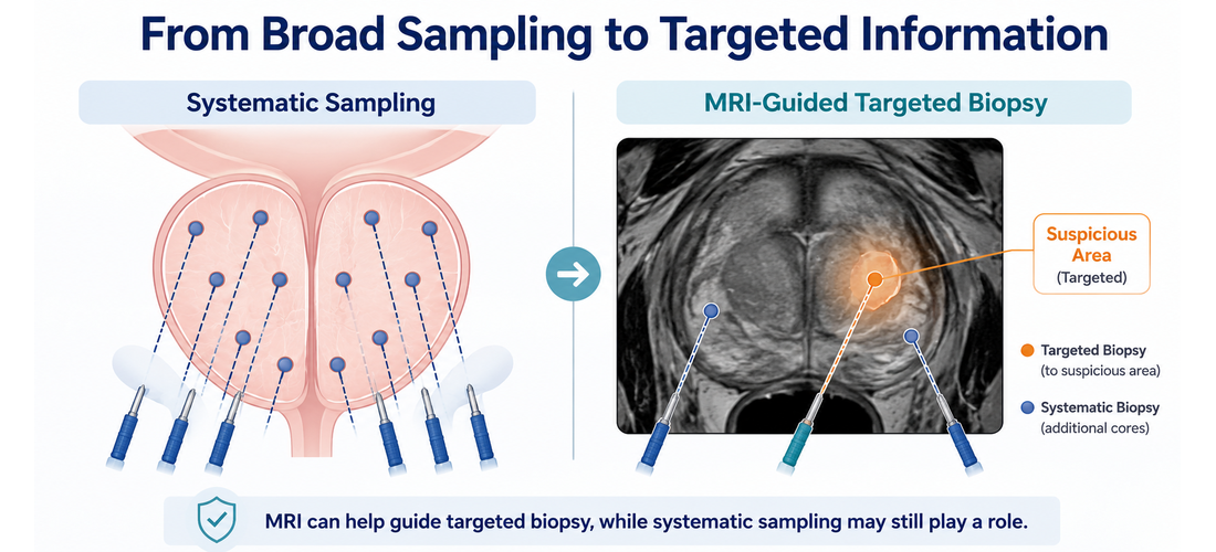
Until relatively recently, most men being evaluated for possible prostate cancer went to biopsy without first having an MRI. Today MRI is often used earlier, although the pathway still varies between patients and health systems.
That variation can be confusing. Two men with similar prostate-specific antigen (PSA) results, measured by a blood test, may receive different advice. One is sent for an MRI first. Another proceeds directly to biopsy, or has additional blood or urine testing first.
None of these pathways is wrong. They reflect improvements in imaging, evolving evidence, regional guidance, local resources, and the fact that prostate cancer risk cannot be judged from one number alone.
For many years, concern rested on a short list of findings: an elevated or rising PSA, an abnormal digital rectal examination (DRE), family history, and symptoms.
When concern was high enough, the next step was usually a systematic biopsy, in which the physician takes tissue samples called cores from a planned pattern across the gland, commonly around 10 to 12.
A prostate cancer diagnosis depends on tissue examined by a pathologist, who determines whether cancer is present and, if it is, assigns a Grade Group reflecting how abnormal and potentially aggressive the cells appear.
Systematic biopsy remains useful, and it has limits. It samples selected areas rather than the whole gland, so a needle can miss a significant tumor, particularly in locations that are harder to reach. It can also find a small, low-risk cancer that may never cause harm, a concern often described as overdiagnosis. Men diagnosed with low-risk disease are frequently candidates for active surveillance, meaning careful monitoring rather than immediate treatment.
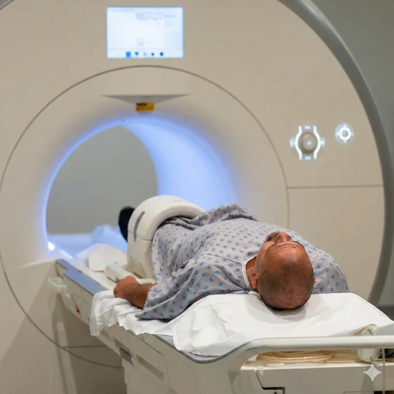
Prostate MRI gives physicians a detailed view of the gland before tissue is removed.
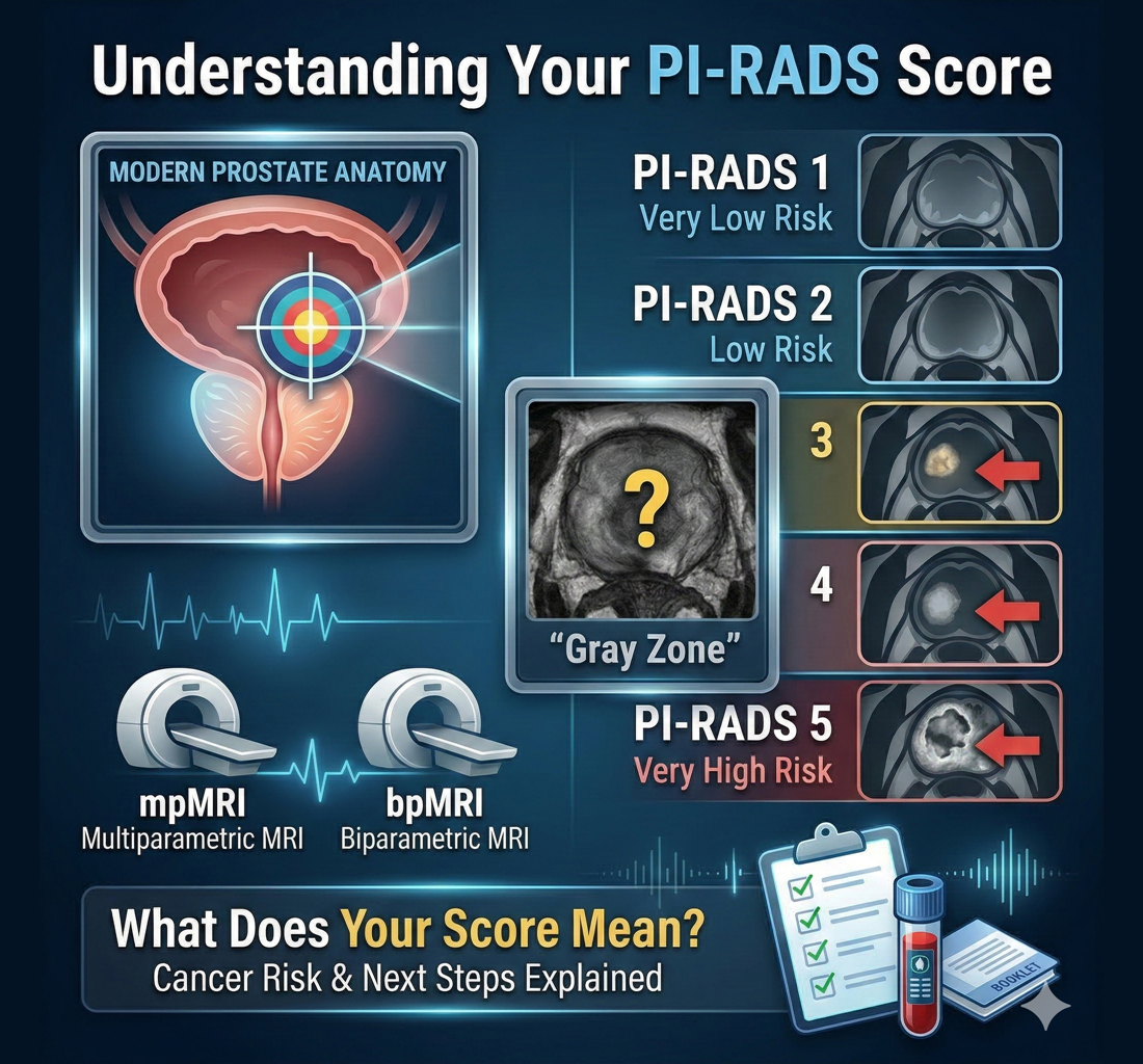
Multiparametric MRI (mpMRI) combines several image types: T2-weighted (T2W) images showing anatomy, diffusion-weighted imaging (DWI) and apparent diffusion coefficient (ADC) maps reflecting how water moves through tissue, and dynamic contrast-enhanced (DCE) images taken after an injected contrast agent. Biparametric MRI (bpMRI) generally omits the contrast sequence.
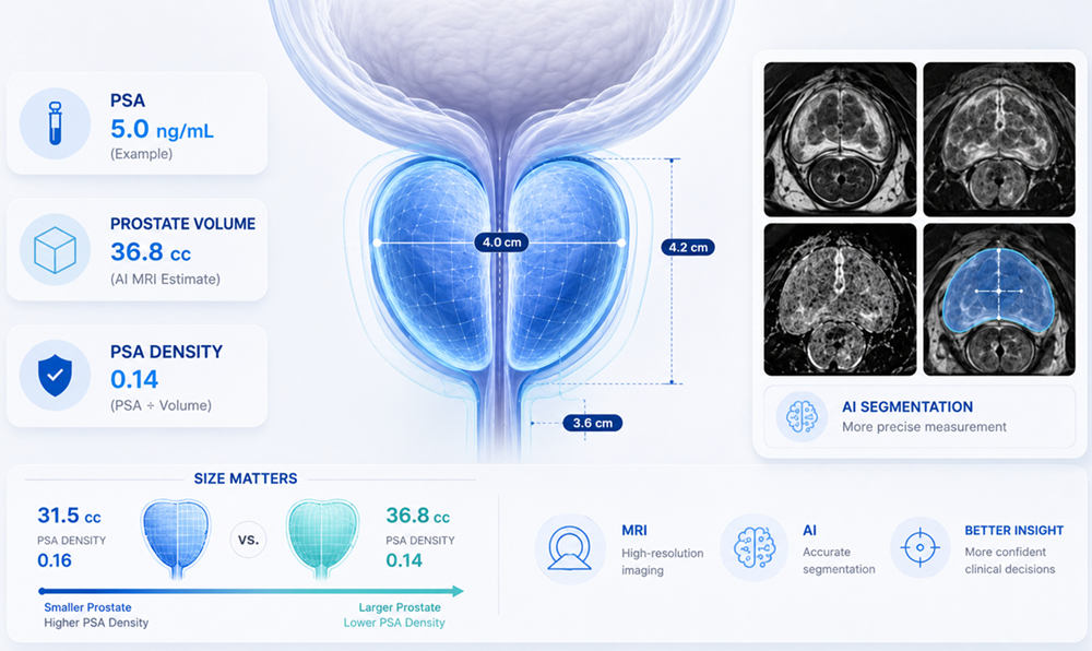
MRI may show suspicious areas and their locations, lesion size and imaging characteristics, prostate volume, and changes related to benign prostatic hyperplasia (BPH, a non-cancerous enlargement common with age).
Radiologists commonly describe how suspicious an area appears using the Prostate Imaging Reporting and Data System (PI-RADS), which runs from 1 to 5.

In the UK, NICE recommends reporting pre-biopsy mpMRI on a 5-point Likert scale, which allows clinical context to be weighed alongside imaging appearance. A PI-RADS or Likert score is not the same as a diagnosis.
MRI has limits in both directions.
Some clinically significant cancers, meaning cancer more likely to affect health or require treatment, are not visible on MRI. Small tumors, diffuse disease, and certain locations can be difficult to see. In the other direction, benign enlargement, inflammation, and other non-cancerous changes can sometimes look suspicious, so a concerning MRI is not confirmation of cancer.
Interpretation varies as well, because scan quality, equipment, lesion size and location, reader experience, and clinical context all matter. Two experienced radiologists reading the same scan do not always assign the same score.
The AUA has also observed that prostate MRI has not yet been shown to improve long-term outcomes such as cancer-specific survival.
When MRI identifies a suspicious area, the physician may direct biopsy needles toward that location. This is called an MRI-targeted biopsy. It may be done by visually matching the MRI to ultrasound images, by using MRI-ultrasound fusion software, or by performing the biopsy inside the MRI scanner.
Major trials, including PROMIS and PRECISION, helped establish MRI's role before an initial biopsy, showing that an MRI-based pathway could improve detection of clinically significant cancer, generally Grade Group 2 or higher, while reducing some unnecessary biopsies and diagnoses of lower-risk disease.
Targeted sampling does not always replace systematic sampling. Research has shown that targeted and systematic approaches each identify cancers the other misses, so depending on MRI findings, prior biopsy history, and overall risk, a physician may take targeted samples alone or combine the two.
A negative MRI lowers risk, but it does not reduce the risk to zero.
In selected patients, an MRI without a suspicious lesion may support a shared decision to defer biopsy and continue PSA monitoring. That decision depends on the full clinical picture, which may include the PSA result and trend, PSA density, prostate volume, PI-RADS or Likert score, age and life expectancy, family history, race or ancestry where clinically relevant, prior biopsy history, symptoms and other conditions, selected biomarkers or inherited genetic findings, and patient preferences.
PSA density can help place a PSA result into context. It divides the PSA level by the prostate volume measured on imaging. Two illustrative examples, both using a PSA of 6.0 ng/mL:
NICE refers to a PSA density above 0.15 ng/mL/cc as one marker of stronger suspicion. These figures are illustrative and should be interpreted by a physician.
Similar PSA and MRI results can therefore mean different things in different men. AUA/SUO guidance notes that when a man remains at elevated risk despite an unremarkable MRI, systematic biopsy is still warranted.
When biopsy is deferred, follow-up matters. A physician may recommend repeat PSA testing, reassessment of PSA density, another MRI, or further evaluation as things change over time.
For more information about prostate size and density, see: What Does Your Prostate Size Actually Tell Your Doctor?
Professional guidance reflects different evidence standards, health systems, and available resources.
Practical factors matter too: MRI access and waiting times, insurance or reimbursement, scan quality, local radiology and biopsy expertise, clinical urgency, and the patient's overall health and preferences.
A different recommendation does not automatically mean that one physician is ignoring evidence. It may reflect a different combination of risk, resources, regional guidance, and clinical circumstances.
AI-supported analysis may add another layer of review by helping identify suspicious imaging patterns, outline the prostate and possible lesions, estimate volume, and highlight regions that may deserve closer attention. How much it improves real-world outcomes is still being studied. It does not confirm cancer or determine whether a biopsy or treatment is needed, and physician interpretation remains central.
For men who already have a prostate MRI, DeepView Imaging provides an AI-supported second analysis powered by ProstatID, AI software from Bot Image that is FDA-cleared, CE-Marked, and UKCA-Certified for prostate MRI analysis. It analyzes T2W, DWI, and ADC images, so it works with both bpMRI and mpMRI scans, and produces a report with color overlays that can be shared with a radiologist, urologist, or treating physician.
This offers another perspective on information already contained in the MRI. It does not replace the original radiology interpretation, biopsy pathology, or physician judgment, and the cleared intended use states that patient-management decisions should not be based solely on the ProstatID analysis.
The growing use of MRI before prostate biopsy reflects better imaging, stronger evidence, and an effort to make biopsy decisions better informed.
MRI can help identify suspicious areas, guide targeted sampling, estimate prostate volume, and place PSA density into context. In some men with a reassuring MRI and low overall risk, it may support a discussion about monitoring rather than immediate biopsy. In others, biopsy may still be appropriate even when MRI does not show a suspicious area.
No single test, including PSA, PSA density, MRI, PI-RADS, biomarkers, or AI-supported analysis, can diagnose or rule out prostate cancer by itself. Your physician can interpret these findings together and explain how they apply to you.
For more perspectives, check out the following articles:
.svg)