September is Prostate Cancer Awareness Month.
Learn the facts and understand your options.
Learn the facts and understand your options.


Your PSA came back higher than expected, your doctor ordered a prostate MRI, and the report came back clean: no suspicious lesion, or a PI-RADS 1 or 2 (in the UK, a Likert score of 1 or 2 on the 5-point scale NICE recommends).
That is real reassurance, and it can still feel unfinished, since the number that started all of this is still elevated. Understanding what the scan was looking for helps explain why "normal MRI" and "let's keep an eye on this" can both be true.
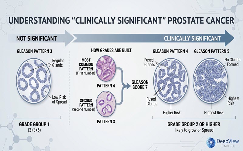
Some prostate cancers grow so slowly that they are unlikely to affect a man's health in his lifetime, and finding them can lead to treatment that causes more harm than the disease would have. Modern prostate care therefore focuses on clinically significant prostate cancer: disease judged likely to grow, spread, or need treatment. In most guidelines that means Grade Group 2 or higher (a Gleason score of 3+4=7 or above). Grade Group 1 disease is often monitored with active surveillance rather than treated right away.
Prostate MRI is built around that target, so it is good at flagging areas that look like significant cancer and less sensitive to small, low-grade disease.
For more information see: What "Clinically Significant" Prostate Cancer Actually Means
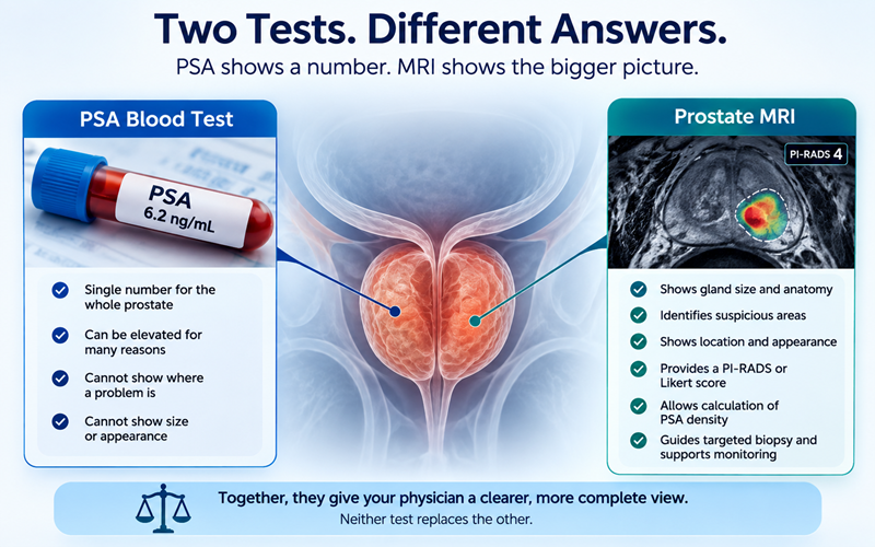
PSA is made by prostate tissue, healthy and cancerous alike. It is prostate-specific rather than cancer-specific, and ordinary things can raise it: benign prostatic hyperplasia (BPH), the age-related enlargement of the prostate; prostatitis or a urinary infection; recent ejaculation, vigorous cycling, a catheter, a cystoscopy, or a recent biopsy; and age itself.
Some medications move it the other way. Finasteride and dutasteride typically cut PSA roughly in half after several months, so a normal-looking result in a man taking them may need to be read differently. A single elevated PSA is often repeated before anyone draws conclusions.
In pooled analyses, the negative predictive value of a good-quality scan is around 90 percent for clinically significant disease, and lower in men whose underlying risk was higher to begin with. A negative MRI lowers risk, but it does not reduce the risk to zero.
Guidelines reflect that balance. In the UK, NICE suggests biopsy may be omitted at a Likert score of 1 or 2 after a shared discussion of risks and benefits. EAU guidance in Europe similarly supports omitting biopsy and continuing PSA monitoring when the MRI is PI-RADS 2 or below and suspicion is otherwise low. In the U.S., AUA/SUO guidance notes some men with a negative MRI may still warrant biopsy when other markers remain concerning.
For more information see: Understanding Your PI-RADS Score
The MRI is one input among several.
Similar numbers can mean different things in different men, which is why two patients with the same PSA are sometimes advised differently.
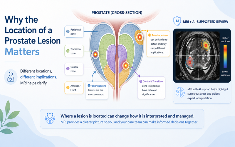
MRI interpretation can vary because scan quality, lesion location, reader experience, and clinical context all matter. A reliable read depends on clear T2-weighted (T2W) images, diffusion-weighted imaging (DWI), and an ADC map, which motion, bowel gas, or an older scanner can degrade. Radiologists grade this formally using PI-QUAL, and lesions in some locations, such as the front of a large prostate, are easier to overlook.
For men who feel reassured but not fully resolved, one option is another look at imaging already done. DeepView Imaging provides an AI-supported second analysis of a prostate MRI using ProstatID™, which is FDA-cleared, CE-Marked, and UKCA-Certified. It reads the T2W, DWI, and ADC data already in the scan and marks regions it scores as suspicious with a color overlay, giving patients and their care team another perspective on information already contained within the MRI.
AI-supported analysis may help highlight areas that deserve closer attention, though physician judgment remains central. DeepView Imaging does not replace a radiologist, urologist, or treating physician, and results can be shared with your doctor when discussing next steps.
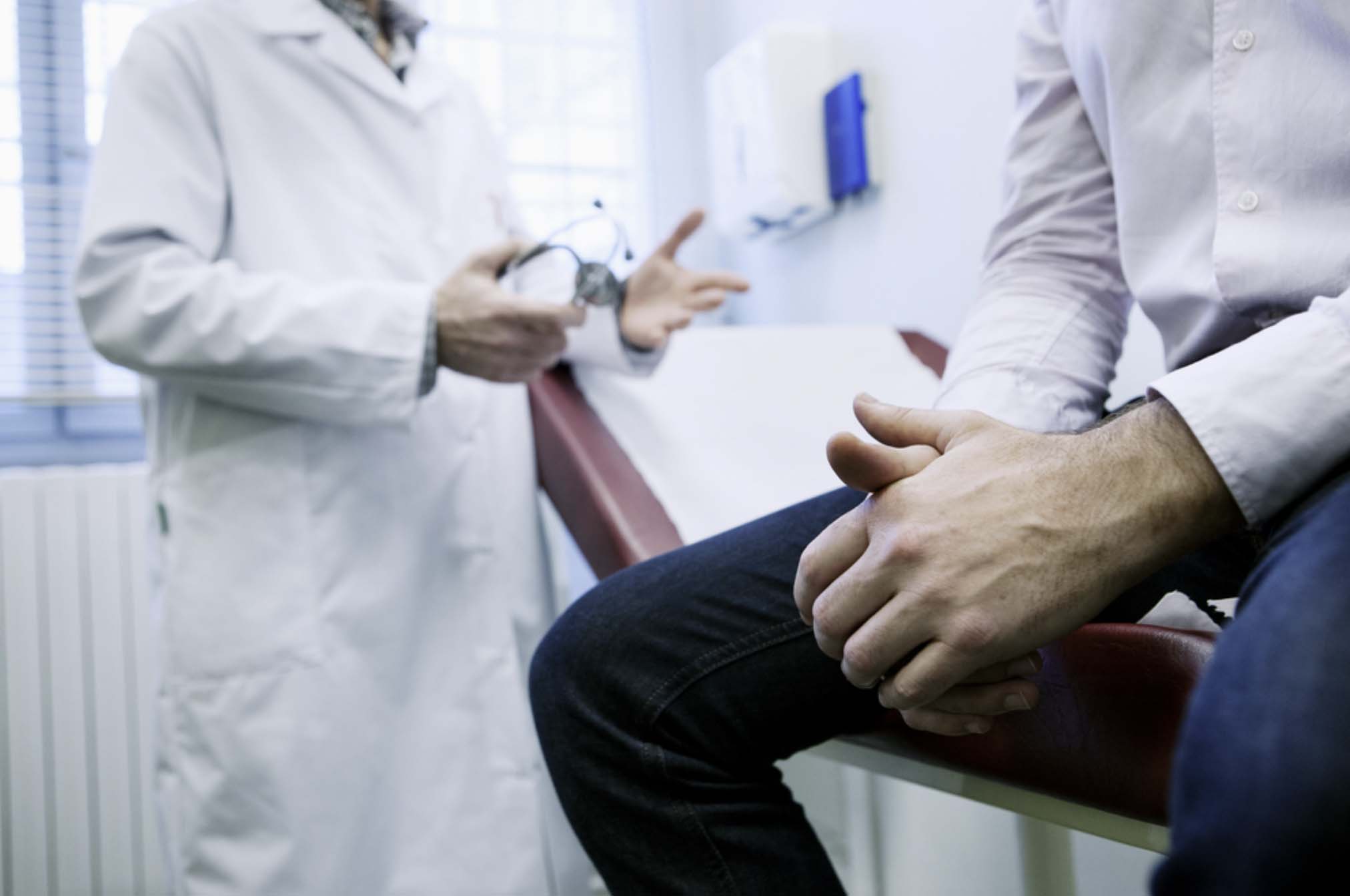
An elevated PSA with a clean MRI is a common and generally reassuring combination. It usually means a shift from urgent evaluation toward careful monitoring, shaped by your PSA trend, PSA density, exam, history, and preferences. A useful question for your next appointment: given what we know so far, what should we watch, how often, and what would change the plan.
Disclaimer:
This article is for educational purposes only and is not medical advice. Decisions about screening, biopsy, diagnosis, monitoring, or treatment should be made with your treating physician.
.svg)