September is Prostate Cancer Awareness Month.
Learn the facts and understand your options.
Learn the facts and understand your options.


Most men leave the imaging center with nothing in hand. A week or two later, a written report arrives, or a physician reads it aloud at an appointment. It is easy to assume that the report is the scan, and that the pictures live somewhere in a hospital system that patients cannot reach.
That assumption costs people time. If you ever want a second opinion, want to switch hospitals, want your surgeon and your oncologist looking at the same study, or simply want a copy of your own for the years ahead, you will need the images themselves. Getting them is usually straightforward once you know who to ask and what to ask for.
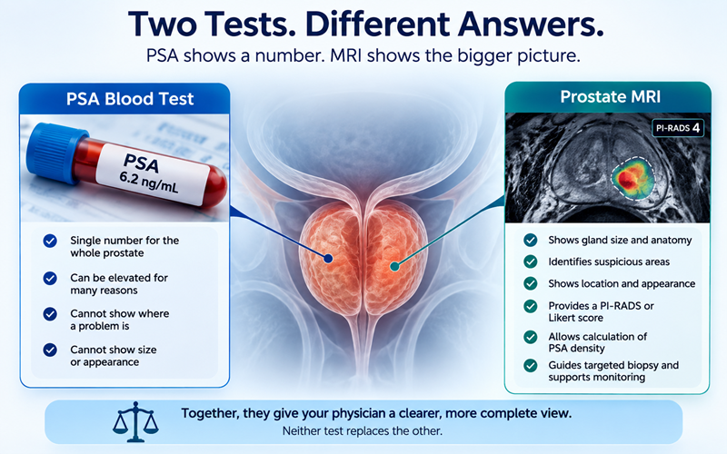
The radiology report is the radiologist's written interpretation. It describes what was seen, often assigns a suspicion score such as PI-RADS (or a Likert score, which is used in parts of the UK), and gives an impression that your physician uses in planning next steps.
The images are the scan itself: hundreds or thousands of individual pictures across several sequences. A prostate MRI typically includes T2-weighted (T2W) images that show anatomy in detail, diffusion-weighted images (DWI) and the matching ADC map, which reflect how water moves within tissue, and sometimes dynamic contrast-enhanced (DCE) images taken after an injection of contrast dye.
Those images are stored in DICOM format, short for Digital Imaging and Communications in Medicine. It is the international standard for medical imaging, and it is what you should ask for by name. A DICOM file is not simply a picture. It also carries the scanner settings, the sequence names, the slice positions, and the information that lets software line one image up with another. Screenshots, JPEGs, and photographs of a screen strip all of that out, which is why they are generally not usable for a formal second review.
A useful way to think about it: the report is the summary someone wrote after reading a book. DICOM is the book. A PDF report cannot be re-read. Images can, and any second review, whether by another radiologist, a tumor board, or an AI-supported analysis, needs the original image data.
Three places hold pieces of your study, and only one of them reliably has the images.
You do not usually need your physician's permission to request your own images.
Being specific prevents most of the common delays. Ask for the complete DICOM study, meaning all series and all sequences rather than selected key images; the radiology report as well; delivery to you, on CD or USB or by secure download; and any prior prostate MRI studies you may have. That last point matters more than people expect, because comparing a current scan to an older one is often one of the most useful things a radiologist can do.
Send it by email if you can, and keep a dated copy.
In the United States, the HIPAA Privacy Rule requires a provider to act on your request within 30 calendar days. One extension of up to 30 more days is permitted, and only if they tell you in writing within the first 30 days why they need it and when they will finish. Fees must be reasonable and cost-based, and federal guidance describes a flat-fee option of up to $6.50 for electronic copies of records held electronically, though this is an option rather than a ceiling on all charges. Some states set shorter deadlines or tighter fee limits. One practical note: fee protections apply most clearly when records go to you. If you ask a facility to send images directly to a third party, different rules and higher charges may apply, so it is often simpler to receive the copy yourself and forward it.
In the EU, Article 15 of the GDPR gives you the right to a copy of your personal data, including imaging. The first copy is free, and the response deadline is one month, extendable by up to two further months for complex or numerous requests, with an explanation.
In the UK, the equivalent right sits under the UK GDPR and the Data Protection Act 2018, with the same one-month deadline and the same free first copy. NHS trusts commonly transfer images through the Image Exchange Portal (IEP) or supply an encrypted disc.
In practice, many centers move faster than the legal maximum. It is not unusual to receive a disc in a few days, particularly if you explain why you need it.
Front-desk staff sometimes say the images "belong to the hospital" or that only a physician can request them. That is usually a misunderstanding of the rules. The facility keeps the original; you have a right to a copy. If you meet resistance, put the request in writing and cite the relevant law as in the letter above, ask for the Privacy Officer (U.S.) or Data Protection Officer (EU/UK) by name, and keep a simple log of dates, names, and promises. If that fails, you can escalate: in the U.S. to the HHS Office for Civil Rights, which has pursued a series of right-of-access enforcement actions; in the UK to the Information Commissioner's Office; and in the EU to your national data protection authority. Most requests never get that far.
Many CDs and USB drives arrive with a small viewer already on them, which is often the easiest starting point. If not, several free programs will open DICOM files:
One caution worth stating plainly. These free viewers are intended for personal review, education, or research, and are generally not cleared for diagnostic use. Opening your own scan can be interesting and can help you follow a conversation with your physician. It is not a substitute for interpretation by someone trained to read prostate MRI, and prostate images in particular are easy to misread without that training.
Having the study in your own hands makes several things easier:
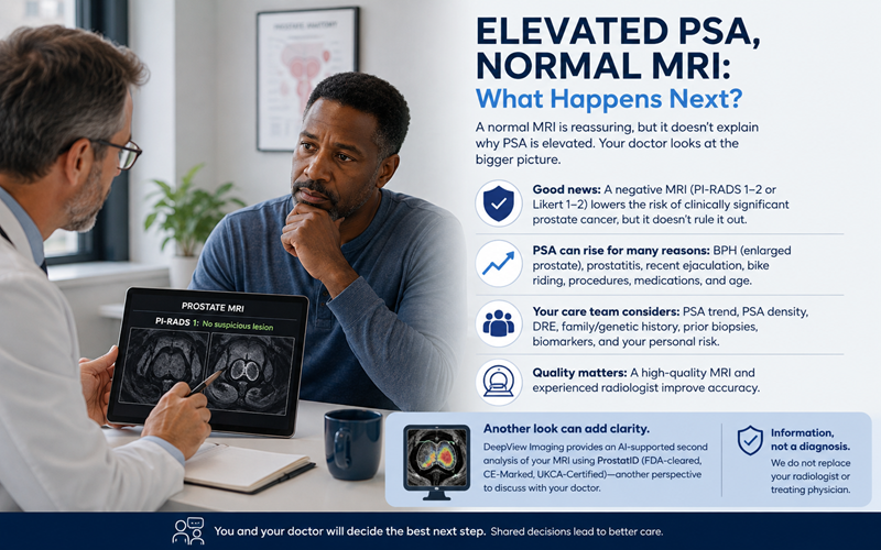
If you already have your MRI in hand, DeepView Imaging provides an AI-supported second analysis of a prostate MRI. A man who has had a scan can securely upload it and receive a report with visual color overlays (on the DICOM images) highlighting regions the software scores as suspicious, which he can then review with his physician. It is powered by ProstatID™, AI software that is FDA-cleared, CE-Marked, and UKCA-Certified for prostate MRI analysis. ProstatID uses the T2-weighted, diffusion-weighted, and ADC data, and does not require the contrast-enhanced (DCE) sequence, so it works with both bpMRI and mpMRI scans. The goal is to give patients and their care team another perspective on information already contained within the MRI. AI-supported analysis may help highlight areas that deserve closer attention, and physician judgment remains central. DeepView Imaging does not replace a radiologist, urologist, or treating physician, and it does not diagnose cancer.
Your prostate MRI report tells you what one radiologist saw. The DICOM images are the underlying record, and you have a legal right to a copy of them in the U.S., the EU, and the UK. Asking for the complete study by name, in writing, addressed to the department that actually stores it, is usually all it takes. Whether you use that copy for a second opinion, for continuity between physicians, or simply to have it on file, having your own images puts you in a stronger position for every conversation that follows.
.svg)