September is Prostate Cancer Awareness Month.
Learn the facts and understand your options.
Learn the facts and understand your options.

Imagine trying to understand a city knowing only its population. The number tells you something real, since a city of 50,000 differs from one of five million. It tells you nothing about the neighbourhoods, the roads, or how the place is actually laid out.
A PSA result works in a broadly similar way. It reports on activity across the gland as a whole. A prostate MRI takes a different approach, letting physicians look at the prostate itself. Understanding that difference explains why many men are offered both.
PSA stands for prostate-specific antigen, a protein made by cells in the prostate. A small amount circulates in the blood, and the test reports how much, usually in nanograms per millilitre (ng/mL).
PSA can rise with prostate cancer. It can also rise for reasons that have nothing to do with cancer, including benign prostatic hyperplasia (BPH, a non-cancerous enlargement common with age), prostatitis (inflammation or infection), a urinary tract infection, recent ejaculation, or certain procedures. An elevated PSA is a reason to look further rather than a diagnosis.
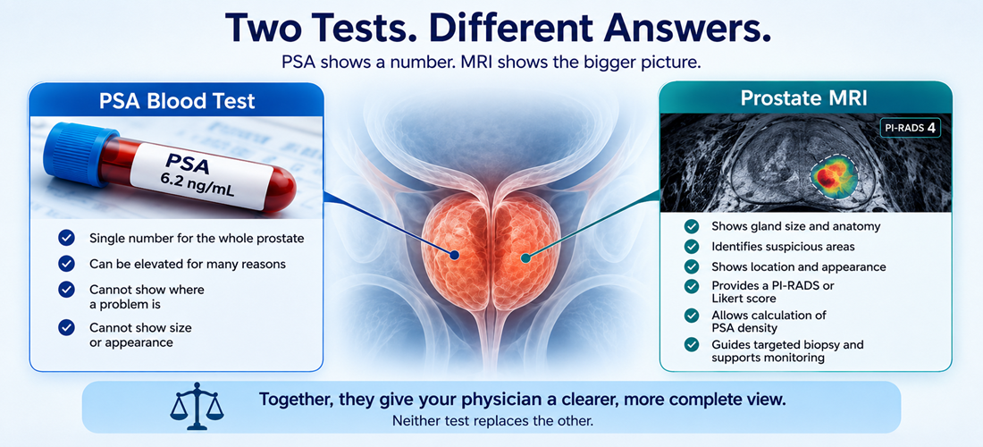
What the number cannot do is point anywhere. It gives no indication of where within the gland a problem might sit, how large it might be, or what it looks like.
An MRI produces detailed images of the prostate and the tissue around it, and this is where the differences become concrete.
It can show where a suspicious area sits, including how close it lies to the edge of the gland. It can show how large that area is, since MRI reports often include lesion measurements. Size alone does not determine whether a lesion is clinically significant, though it contributes context during risk assessment. Imaging can also reveal more than one area of interest, or asymmetry between the two sides.
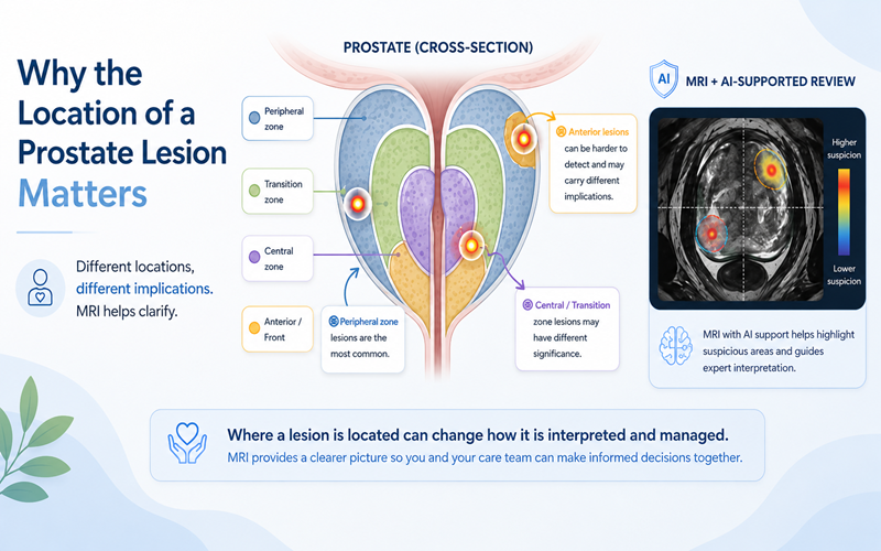
Location matters because the prostate is not uniform tissue. It contains distinct zones, and the two most often discussed are the peripheral zone, where most clinically significant cancers arise, and the transition zone, where benign enlargement tends to occur. A finding in one zone may carry different weight from a similar finding in another. A blood test cannot make that distinction.
Different MRI sequences reveal different things. T2-weighted (T2W) images show anatomy. Diffusion-weighted imaging (DWI) and the ADC map reflect how freely water moves through tissue, which relates to how tightly packed the cells appear. Multiparametric MRI (mpMRI) adds dynamic contrast-enhanced (DCE) images; biparametric MRI (bpMRI) omits that contrast sequence.
Radiologists summarize these observations as a suspicion score. PI-RADS, a 1 to 5 scale from the ACR and ESUR, is used across the U.S. and much of Europe. In the UK, NICE guidance also supports a 5-point Likert scale. Both describe how suspicious an area appears on imaging, and neither is the same as a diagnosis.

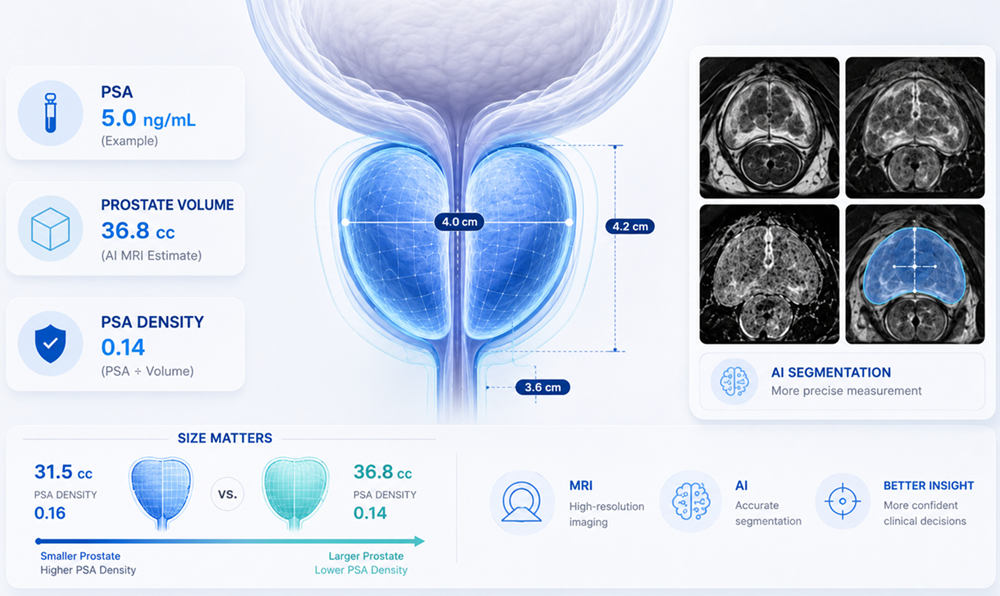
Because MRI enables the gland's size to be measured, it allows PSA density to be calculated: PSA divided by prostate volume.
Consider two illustrative men, each with a PSA of 6 ng/mL. One has a 30 cc prostate, giving 6 ÷ 30 = 0.20. The other has a 90 cc prostate, giving 6 ÷ 90 = about 0.07. The same blood result carries different weight in each case. These are examples only, and the thresholds physicians apply vary, so any real number should be interpreted by your care team.
Measuring the size of the prostate is accomplished in one of three ways:
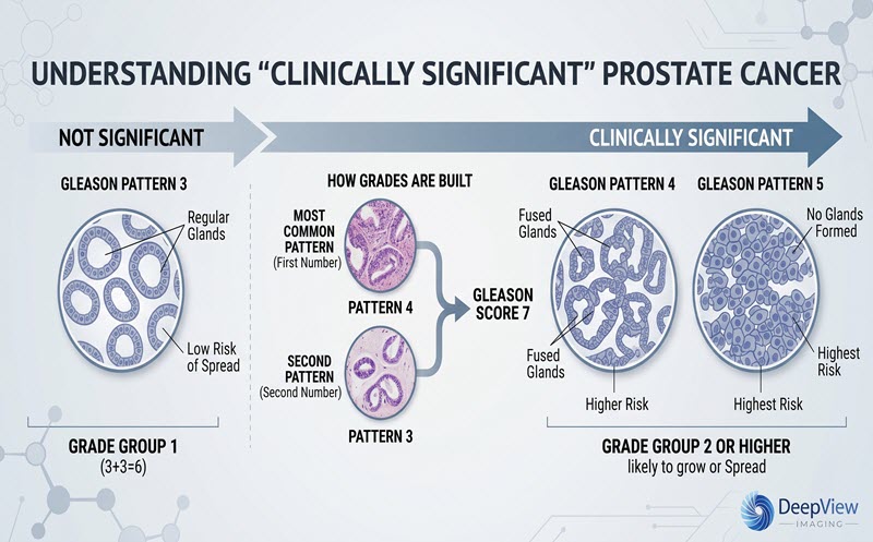
MRI does not confirm cancer. Diagnosis generally depends on biopsy and pathology, which is also where a Gleason score and Grade Group come from.
MRI can miss clinically significant prostate cancer. A negative MRI lowers risk, though it does not reduce the risk to zero. Interpretation can vary as well, because scan quality, lesion location, reader experience, and clinical context all matter.
Decisions therefore rest on the whole picture: your PSA trend over time, PSA density, MRI findings and score, family history, age, ancestry where clinically relevant, prior biopsy history, symptoms, other health conditions, and your own preferences. Similar numbers can mean different things in different patients. Screening itself is framed as a shared decision under USPSTF guidance in the U.S., as a risk-adapted discussion in EAU guidance across Europe, and through NICE pathways in the UK.
There is one further difference worth knowing. A PSA result is a single value at a single moment. An MRI generates a large volume of imaging data that can be reviewed, measured, and revisited long after the appointment ends. Subspecialist radiologists may offer a second reading. Later scans can be compared against it during active surveillance. AI-supported software may examine measurements and imaging patterns that are difficult to assess by eye. Performance across different scanners and populations remains an active area of study, and physician judgment remains central.
DeepView Imaging provides an AI-supported second analysis of a prostate MRI using ProstatID, which is FDA-cleared, CE-Marked, and UKCA-Certified. ProstatID analyses bpMRI data (T2W, DWI, and ADC) and presents its findings as a color overlay with a risk score, so it works with both bpMRI and mpMRI scans. The goal is to give patients and their care team another perspective on information already contained within the MRI. DeepView Imaging does not replace a radiologist, urologist, or treating physician, and patients can share their results with their physician when discussing next steps.
A PSA test suggests that something may warrant a closer look. An MRI helps show where to look, how large the area is, and how concerning it appears. Together they give your physician a fuller picture, and understanding the difference puts you in a stronger position to ask good questions about what comes next.
This article is for educational purposes only and is not medical advice. Decisions about screening, biopsy, diagnosis, monitoring, or treatment should be made with your treating physician.
.svg)