Rising PSA After a Negative Biopsy: What Are Your Options?

You had the biopsy. The pathology report came back benign. For a moment, it felt like the story was over.
Then your next PSA test came back higher. Or the one after that. Now you are left with a confusing question: If the biopsy found no cancer, why is my PSA still rising?
There are several possible explanations, many unrelated to cancer. A negative biopsy is reassuring, but it cannot examine every part of the prostate. Understanding what your tests did and did not show can help you discuss your next steps.
Why can a negative biopsy still leave questions?
A negative biopsy is good news. It provides some reassurance, and U.S. guidelines advise physicians to account for that result when estimating future cancer risk.
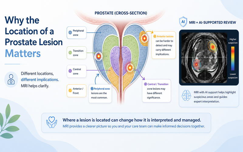
Still, a biopsy examines only the tissue collected by its needles. A systematic biopsy typically takes about 10 to 12 small samples, called cores, from different areas of the prostate. It can be performed through the rectum or through the skin between the scrotum and anus. Small or difficult-to-reach areas, such as the front (anterior) part of the prostate, may remain unsampled.
The UK PROMIS study, published in The Lancet in 2017, compared prostate MRI and standard transrectal biopsy with a more extensive mapping biopsy. MRI identified 93% of cancers meeting the study's definition of clinically significant disease, while standard transrectal biopsy identified 48%. MRI also flagged some harmless areas. Importantly, the men were undergoing their first biopsy, so these findings cannot predict an individual's repeat-biopsy results.
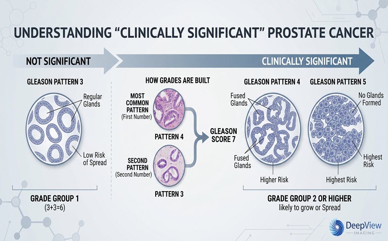
Clinically significant prostate cancer generally means cancer more likely to grow or cause harm, often defined in research as Grade Group 2 or higher. Some lower-risk cancers grow slowly and may not need immediate treatment.
A rising PSA may lead your physician to reassess your risk, but it does not prove cancer was missed.
For more information see: Why Lesion Location Matters
Not necessarily. PSA (prostate-specific antigen) is produced by normal as well as cancerous prostate tissue. PSA may rise because of:
- Benign prostatic hyperplasia (BPH): Noncancerous enlargement of the prostate.
- Prostatitis or infection: Inflammation or infection that may temporarily raise PSA.
- Recent prostate procedures: A biopsy can affect PSA for a period afterward.
- Short-term factors: Ejaculation and, in some circumstances, vigorous cycling.
Your physician may consider the timing of your tests, temporary factors, and the PSA trend rather than relying on a single number.
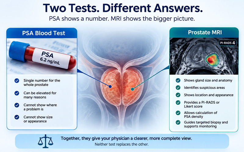
PSA density helps place the result into context by dividing PSA by prostate volume, often measured on MRI. For example, a PSA of 7.2 ng/mL in an 80 mL prostate gives a density of 0.09; the same PSA in a 30 mL prostate gives 0.24. A larger prostate can produce more PSA without cancer.
Higher PSA density can increase concern, but no single density value rules cancer in or out. MRI findings, family history, and other factors remain important. Medications such as finasteride and dutasteride also matter because they can substantially lower measured PSA.
For more information see: Why Prostate Size Matters
If you have not had a prostate MRI, your physician may discuss one before a repeat biopsy. The American Urological Association/Society of Urologic Oncology (AUA/SUO) guideline, updated in 2026, recommends MRI before repeat biopsy when no previous prostate MRI is available.
MRI can show suspicious regions, guide targeted biopsy, and support monitoring discussions. But it cannot diagnose cancer by itself. A negative MRI lowers risk, but it does not reduce the risk to zero.
Radiologists commonly use PI-RADS, a five-point scale describing how suspicious an area appears. In the UK, NICE guidance uses a similar five-point Likert scale. Neither score confirms cancer.
If you already had an MRI, two separate questions matter:
Was the scan technically adequate? Motion, rectal gas, implants, or acquisition problems can reduce image quality. PI-QUAL, updated in 2024, is a three-point system for assessing MRI quality, including scans with or without contrast. A scan with major limitations may need to be repeated.
How was the scan interpreted? Even good-quality images may be read differently depending on the radiologist's experience and the location or appearance of a suspicious area. Review by an experienced prostate radiologist may add another perspective.
A second interpretation cannot recover information that was never captured clearly in the first scan.
For more information see: Elevated PSA, Normal MRI: What Happens Next? and Why MRI Interpretation can vary
Two choices shape a biopsy: where the samples are taken and which route the needles use.
Targeted versus systematic biopsy. A targeted biopsy samples a suspicious MRI region, sometimes using MRI-ultrasound fusion. A systematic biopsy samples several planned areas. Depending on the situation, a physician may use one approach or combine them. A systematic repeat biopsy may still be considered when MRI is negative but clinical concern remains.
Transperineal versus transrectal biopsy. A transperineal needle passes through skin between the scrotum and anus. A transrectal needle passes through the rectal wall. Either route can be used for targeted or systematic sampling.
Two randomized trials compared these routes in men having their first, rather than a repeat, biopsy:
What does this mean for patients? Transperineal biopsy avoids passing the needle through the rectal wall, generally lowers infection risk, and may help reach certain areas. It can involve more immediate discomfort or a longer procedure, and temporary difficulty urinating can occur. Transrectal biopsy remains an option with appropriate infection precautions. European guidance favors transperineal biopsy; U.S. guidance allows either, and NICE supports local-anesthetic transperineal biopsy in the UK.
Because both trials studied first biopsies, their percentages should not be treated as your personal chances after a previous negative biopsy.
For more information see: Understanding Prostate Biopsy Decisions
Some tests may refine your risk estimate when it is unclear whether another biopsy would be useful:
- Blood biomarkers: Such as Prostate Health Index (PHI), 4Kscore, and Stockholm3.
- Urine biomarkers: Such as MyProstateScore, ExoDx, and PCA3-based testing.
- Tissue-based tests: Tests that analyze samples from an earlier negative biopsy for molecular changes associated with risk.
These tests have different evidence, intended uses, and availability. In the U.S., AUA/SUO guidance supports selective use after a negative biopsy when a result could change the next decision, rather than routine testing for everyone.
European guidance also considers biomarkers in some settings. NICE, however, does not recommend the PROGENSA PCA3 assay or PHI for people with a negative or inconclusive biopsy. No biomarker alone can confirm or exclude cancer.
AUA/SUO guidance advises against using a PSA cutoff alone to decide on repeat biopsy. Your team may consider PSA trend and density, prostate volume, MRI findings and quality, PI-RADS or Likert score, lesion location, prior biopsy details, age, family history, ancestry, symptoms, other medical conditions, and your preferences.
Depending on the clinical picture, monitoring, additional MRI review, selected biomarkers, or repeat biopsy may be discussed. If low-risk cancer is eventually found, active surveillance (regular monitoring instead of immediate treatment) may be suitable for some patients.
Five questions to ask your urologist
- Could something other than cancer explain my rising PSA?
- Did the first biopsy sample suspicious areas seen on MRI?
- Was my MRI good enough, and might an experienced second reader or repeat scan help?
- Would PSA density or a selected biomarker test help clarify my risk?
- What are the benefits and risks of monitoring versus another biopsy in my situation?
DeepView Imaging offers an AI-supported second analysis of an existing prostate MRI using ProstatID, an FDA-cleared, CE-Marked, and UKCA-Certified tool designed to assist physicians. Its report includes color overlays and risk scores highlighting regions that may deserve closer attention.
According to Bot Image, ProstatID was trained using data from 1.5T and 3.0T MRI scanners and supports compatible systems from several manufacturers. It also checks for required image sequences and technical or image-quality problems relevant to its acquisition and processing requirements. These checks can help identify scans unsuitable for analysis, but they do not establish that every clinical image-quality issue has been detected or replace a radiologist's assessment. ProstatID analyzes T2-weighted, diffusion-weighted, and ADC images, so compatible scans with or without contrast can be processed.
AI-supported review cannot fix poor-quality images, diagnose cancer, or determine whether another biopsy is needed. DeepView's results are an additional perspective to share with your radiologist, urologist, or treating physician.
For more information see: How to Get a Copy of Your Own Prostate MRI
A negative biopsy is reassuring, but a PSA that keeps rising deserves thoughtful follow-up. There may be a benign explanation, or an area may need closer evaluation.
Understanding your original biopsy, MRI quality and interpretation, and possible next tests can make your discussion with your physician more productive. The appropriate next step depends on the full clinical picture, not one number or test alone.
This article is for educational purposes only and is not medical advice. Decisions about screening, biopsy, diagnosis, monitoring, or treatment should be made with your treating physician.
Sources
- American Urological Association and Society of Urologic Oncology. Early Detection of Prostate Cancer: AUA/SUO Guideline, 2023, amended 2026; 2026 update.
- European Association of Urology. EAU Guidelines on Prostate Cancer: Diagnostic Evaluation, 2026.
- Ahmed HU, et al. Diagnostic accuracy of multi-parametric MRI and TRUS biopsy in prostate cancer (PROMIS). The Lancet. 2017;389:815–822.
- National Cancer Institute. Prostate-Specific Antigen (PSA) Test.
- de Rooij M, et al. PI-QUAL version 2: an update of a standardized scoring system for the assessment of image quality of prostate MRI. European Radiology. 2024;34:7068–7079.
- Hu JC, et al. Transperineal vs. Transrectal Prostate Biopsy: The PREVENT Randomized Clinical Trial. JAMA Oncology. 2024.
- Bryant RJ, et al. Local anesthetic transperineal biopsy versus transrectal biopsy (TRANSLATE). The Lancet Oncology. 2025;26:583–595.
- NICE. Prostate cancer: diagnosis and management (NG131); Transperineal biopsy for diagnosing prostate cancer (HTG680).
- U.S. Food and Drug Administration. ProstatID 510(k) clearance (K212783); FDA indications and technical summary.
- Bot Image. ProstatID training and scanner information; Image intake and quality-check description. Manufacturer descriptions of product capabilities.
- NICE. AI technologies to help detect prostate cancer on MRI: final scope, March 2026 (lists ProstatID as UKCA-marked).
More Articles