Prostate MRI vs. Biopsy: Accuracy, Limitations, and What Results Mean

Your prostate MRI showed a suspicious area, but your biopsy was negative. Or perhaps your biopsy found cancer where the MRI showed little concern. Which result should you trust?
When a biopsy finds cancer, you can generally rely on cancer being present, although the grade it assigns can change with further sampling. A negative biopsy is less certain than many men assume, because it only reflects the samples taken. MRI has limits of its own, and understanding both may help you discuss the results with your doctor.
What can an MRI and a biopsy each tell you?
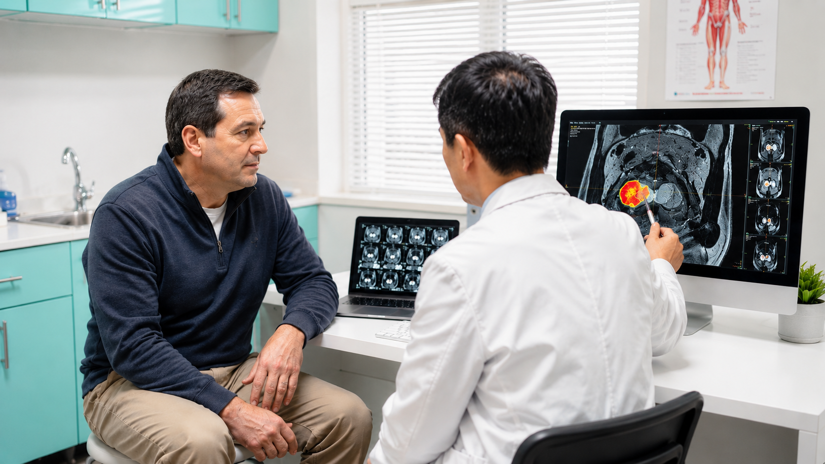
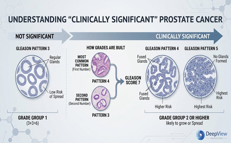
A prostate MRI shows detailed images of the gland. A radiologist looks for clinically significant prostate cancer, often defined as Grade Group 2 or higher on a 1-to-5 scale of how abnormal the cells look under a microscope. Such cancers may need closer attention, but grade alone does not determine treatment.
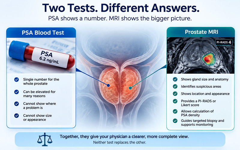
Suspicious areas receive a score from 1 to 5, usually using PI-RADS (Prostate Imaging Reporting and Data System). UK guidance uses a similar Likert scale. Higher scores mean greater suspicion, not a diagnosis. How to Read Your Prostate MRI Results walks through the report in more detail.
A prostate biopsy removes small tissue samples for a pathologist to examine. It can confirm cancer and establish the Gleason score and Grade Group.
A systematic biopsy samples several regions in a planned pattern. An MRI-targeted biopsy samples an area highlighted on the scan. Biopsy needles may pass through the rectum (transrectal) or through the skin between the scrotum and anus (transperineal).
MRI can guide biopsy and support follow-up. Biopsy generally provides the tissue evidence needed for diagnosis. Researchers are studying whether a very confident MRI read might one day carry more weight for some men, though biopsy remains the standard today.
In the UK PROMIS study, 576 men underwent MRI, a standard biopsy, and a more extensive mapping biopsy (many samples taken across the whole gland) used for comparison. MRI identified 93% of cancers that met the study's definition of clinically significant disease. Among men with a negative MRI, 89% did not have cancer meeting that definition. A standard biopsy without MRI targeting identified 48% of those significant cancers.
These figures reflect the study's own definition and patients. MRI accuracy can vary, and they do not represent an individual's chance of having cancer.
The MRI score is important, too. A 2024 review of 70 studies reported these average rates of clinically significant cancer found on biopsy:
These are averages. Results vary with MRI quality, lesion location, patients studied, and reader experience.
Nearly half of PI-RADS 4 findings in these studies were not clinically significant cancer. And a negative MRI lowers risk, but it does not reduce the risk to zero. Can a Prostate MRI Miss Cancer? looks at why.
Yes. A biopsy samples only a small amount of tissue. A negative result means cancer was not found in those samples, and cancer elsewhere in the gland may still be present.
The international PRECISION trial studied 500 men who had not previously undergone biopsy. Clinically significant cancer was detected in 38% of men assigned to MRI followed by targeted biopsy when indicated, compared with 26% assigned to standard biopsy. Low-grade cancer was diagnosed less often in the MRI pathway (9% versus 22%). Under the study's MRI-first protocol, 28% of men did not undergo biopsy because their MRI was not suspicious. This does not mean that biopsy can safely be skipped for everyone with a negative MRI. Results like these help explain why more patients are getting an MRI before their first biopsy.
Biopsies sometimes underestimate cancer grade. In one audit of nearly 3,000 men, the grade found after prostate-removal surgery was higher than the biopsy grade in roughly 3 in 10. These men all went on to surgery, so the finding may not apply to every patient. A 2020 review found that MRI-targeted samples were less likely than systematic samples to underestimate grade.
The biopsy route can differ as well. In Europe, EAU guidance strongly recommends the transperineal route because of its lower risk of infection. In the U.S., AUA/SUO guidance allows either route.
Different results do not necessarily mean that either test was wrong:
- Suspicious MRI, negative biopsy: The MRI finding may reflect inflammation or benign prostatic hyperplasia (BPH), a noncancerous enlargement of the prostate. It is also possible that the needle did not sample the suspicious tissue. Depending on how suspicious the area looked, physicians may consider a second review of the images and biopsy slides, PSA monitoring, a repeat MRI, or a repeat biopsy with targeted samples. Rising PSA After a Negative Biopsy covers these options in more detail.
- Negative MRI, rising PSA: MRI may miss some cancers. Your physician may consider your PSA density (PSA level divided by prostate volume), changes in PSA over time, and other risk factors, as discussed in Elevated PSA, Normal MRI: What Happens Next?
- Low-grade cancer on biopsy, more suspicious MRI: The MRI may raise questions about whether the biopsy captured the most concerning area. Additional review or sampling may be considered before deciding whether active surveillance (careful monitoring instead of immediate treatment) is suitable.
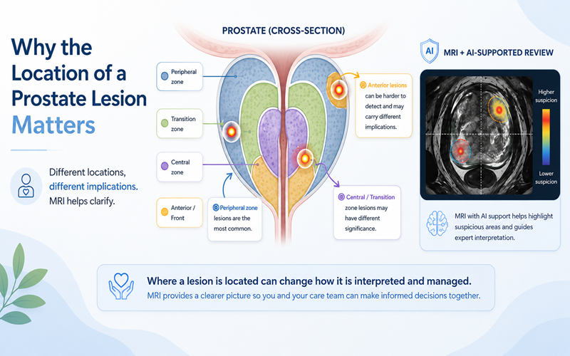
Biopsy decisions often depend on the full clinical picture. Your physician also considers PSA trend, PSA density, prostate size, MRI score and lesion location, age, family history, ancestry when relevant, prior biopsies, symptoms, overall health, and your preferences. Similar results can mean different things for different people.
Questions you may want to ask your physician include:
- How suspicious was the MRI finding, and where was it located?
- Were MRI-targeted, systematic, or both types of samples taken?
- How does my PSA density affect the interpretation?
- Would a review of the MRI images or biopsy slides help explain the results?
The MRI-FIRST study showed that targeted and systematic biopsies each detected some significant cancers that the other missed. More recently, a 2026 study of 902 men with PI-RADS 3 findings found that targeted biopsy alone missed 59 significant cancers detected when systematic samples were also taken. That represented about one-quarter of the significant cancers found with combined sampling, or approximately 6.5% of all men studied.
Extra samples may add less for some higher-suspicion findings. In a 2026 retrospective study of 470 men, systematic samples found additional significant cancer in only about 2 in 100 men overall, and none in men with a PI-RADS 5 finding and a PSA density above 0.20. That result needs confirmation in larger studies.
Guidelines therefore allow for differences in approach. In the United States, AUA/SUO guidance, amended in 2026, recommends targeting suspicious lesions and allows systematic samples to be added. European EAU guidance recommends targeted biopsy with additional samples taken near the suspicious area for PI-RADS 4 or 5 findings, while decisions about PI-RADS 3 findings depend on other risk factors. In the UK, NICE recommends MRI first when cancer is suspected and appears confined to the prostate, then a biopsy guided by the MRI findings for Likert scores of 3 or higher. Decisions should reflect individual risk and shared discussion.
MRI interpretation can vary with scan quality, the location of a suspicious area, and the reader's experience. A 2022 review found substantial agreement between radiologists overall, with more disagreement around borderline findings such as PI-RADS 3. When MRI and biopsy results disagree, another look at the images may highlight details worth discussing.
DeepView Imaging provides an AI-supported second analysis of a prostate MRI you have already had, using ProstatID, software that is FDA-cleared, CE-Marked, and UKCA-Certified. The report marks suspicious regions with visual overlays and risk scores. It analyzes T2-weighted and diffusion-weighted images, so it works with scans done with or without contrast. The goal is to give patients and their care team another perspective on information already contained within the MRI, and patients can share the report with their physician when discussing next steps.
If the AI analysis differs from your original report, that does not automatically mean cancer was missed. It is something to discuss with your physician in context. DeepView Imaging does not diagnose cancer, and it does not replace biopsy, pathology, or a radiologist, urologist, or treating physician. Why training data matters explains how tools like this are built and tested.
So which result should you trust? Each one, for the question it answers. MRI can show where clinically significant prostate cancer may be present. Biopsy examines tissue to confirm cancer and assess its grade. Both have limits, and a disagreement between them is a reason for a closer look. Discuss with your physician whether monitoring, another review, or further testing makes sense for you.
Sources
- Ahmed HU, et al. PROMIS: Diagnostic accuracy of multiparametric MRI and TRUS biopsy. The Lancet. 2017.
- Oerther B, et al. Update on PI-RADS Version 2.1 Diagnostic Performance Benchmarks. Radiology. 2024.
- Kasivisvanathan V, et al. MRI-Targeted or Standard Biopsy for Prostate-Cancer Diagnosis (PRECISION). New England Journal of Medicine. 2018.
- Stapleton P, Milton T, et al. The Effect of Pre-Biopsy Prostate MRI on the Congruency and Upgrading of Gleason Grade Groups Between Prostate Biopsy and Radical Prostatectomy. Société Internationale d'Urologie Journal. 2024.
- Goel S, et al. Concordance Between Biopsy and Radical Prostatectomy Pathology. European Urology Oncology. 2020.
- Rouvière O, et al. MRI-FIRST: Systematic and targeted biopsy. The Lancet Oncology. 2019.
- Buhas BA, Mjaess G, Diamand R, et al. Risk-adapted omission of systematic cores in PI-RADS 3 lesions. European Urology Oncology. 2026.
- Tiro J, et al. Are template prostate biopsies still required in patients with high-risk PI-RADS lesions? A retrospective study of 470 patients. BJUI Compass. October 2026.
- Lin DW, Carlsson S, Filson CP, et al. Updates to Early Detection of Prostate Cancer: AUA/SUO Guideline (2026). The Journal of Urology. 2026.
- European Association of Urology. 2026 Guidelines: Diagnostic Evaluation.
- National Institute for Health and Care Excellence. NG131: Prostate Cancer, Diagnosis and Management.
- Wen J, Ji Y, Han J, Shen X, Qiu Y. Inter-reader agreement of PI-RADS v2.1: A systematic review and meta-analysis. Frontiers in Oncology. 2022.
- U.S. Food and Drug Administration. ProstatID 510(k) K212783. 2022.
- Bot Image, Inc. CE marking announcement (2025); UKCA certification announcement (2025). Manufacturer statements about regulatory status.
More Articles